

Simulating clinical features on chest radiographs for medical image exploration and CNN explainability using a style-based generative adversarial autoencoder
- PMID: 39424900
- PMCID: PMC11489724
- DOI: 10.1038/s41598-024-75886-0
Simulating clinical features on chest radiographs for medical image exploration and CNN explainability using a style-based generative adversarial autoencoder
Abstract
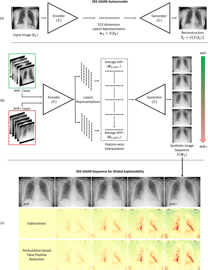
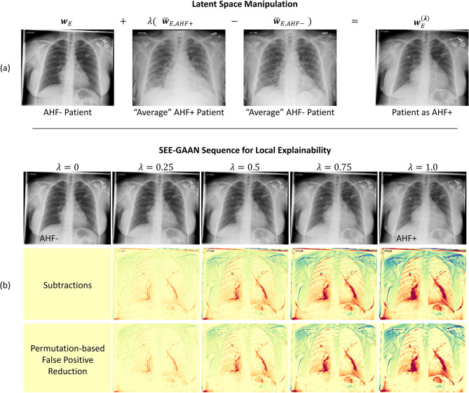
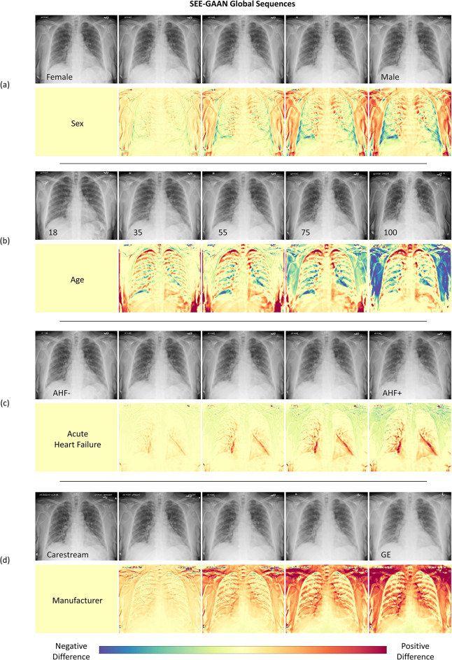
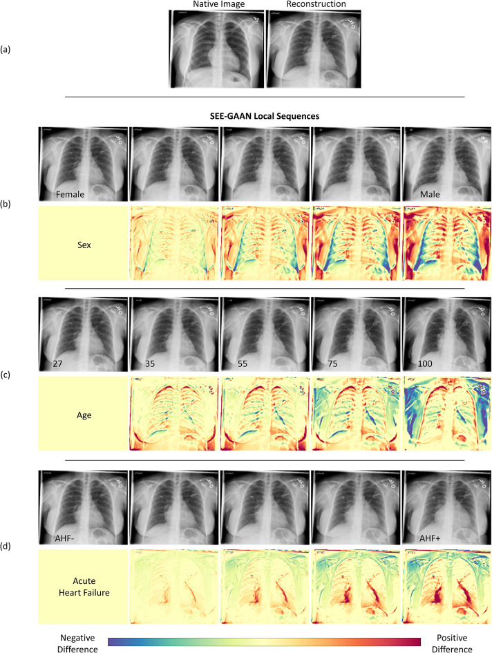
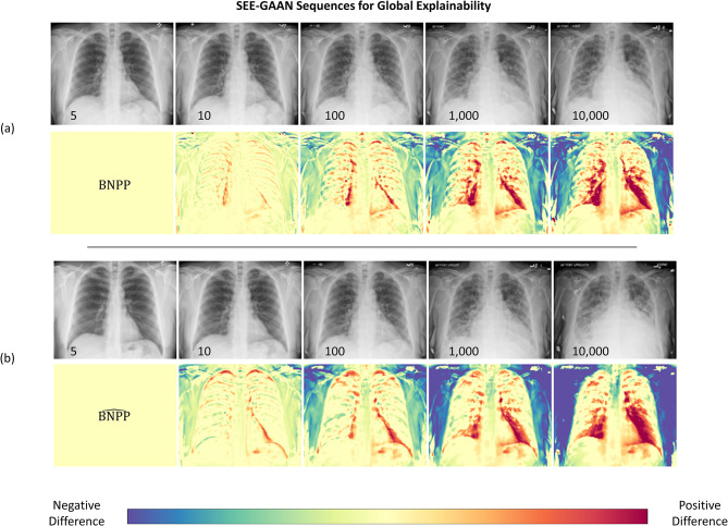
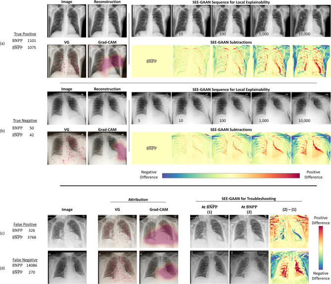
Explainability of convolutional neural networks (CNNs) is integral for their adoption into radiological practice. Commonly used attribution methods localize image areas important for CNN prediction but do not characterize relevant imaging features underlying these areas, acting as a barrier to the adoption of CNNs for clinical use. We therefore propose Semantic Exploration and Explainability using a Style-based Generative Adversarial Autoencoder Network (SEE-GAAN), an explainability framework that uses latent space manipulation to generate a sequence of synthetic images that semantically visualizes how clinical and CNN features manifest within medical images. Visual analysis of changes in these sequences then facilitates the interpretation of features, thereby improving explainability. SEE-GAAN was first developed on a cohort of 26,664 chest radiographs across 15,409 patients from our institution. SEE-GAAN sequences were then generated across several clinical features and CNN predictions of NT-pro B-type natriuretic peptide (BNPP) as a proxy for acute heart failure. Radiological interpretations indicated SEE-GAAN sequences captured relevant changes in anatomical and pathological morphology associated with clinical and CNN predictions and clarified ambiguous areas highlighted by commonly used attribution methods. Our study demonstrates SEE-GAAN can facilitate our understanding of clinical features for imaging biomarker exploration and improve CNN transparency over commonly used explainability methods.
Keywords: Autoencoder; Chest radiographs; Convolutional neural network; Explainable artificial intelligence; Generative adversarial network.
© 2024. The Author(s).
Conflict of interest statement
Dr. Hasenstab, Dr. Hahn, and Mr. Chao declare no potential conflicts of interest. Dr. Hsiao receives research grant support from GE healthcare, Bayer AG, and Bracco. He is a consultant for Canon and was a cofounder of Arterys Inc, which has been acquired by Tempus AI.
Figures
Similar articles
-
Chest Radiographs in Congestive Heart Failure: Visualizing Neural Network Learning.Radiology. 2019 Feb;290(2):514-522. doi: 10.1148/radiol.2018180887. Epub 2018 Nov 6. Radiology. 2019. PMID: 30398431
-
EEG-Based Feature Classification Combining 3D-Convolutional Neural Networks with Generative Adversarial Networks for Motor Imagery.J Integr Neurosci. 2024 Aug 20;23(8):153. doi: 10.31083/j.jin2308153. J Integr Neurosci. 2024. PMID: 39207066
-
Assessment of Convolutional Neural Networks for Automated Classification of Chest Radiographs.Radiology. 2019 Feb;290(2):537-544. doi: 10.1148/radiol.2018181422. Epub 2018 Nov 13. Radiology. 2019. PMID: 30422093 Free PMC article.
-
From CNNs to GANs for cross-modality medical image estimation.Comput Biol Med. 2022 Jul;146:105556. doi: 10.1016/j.compbiomed.2022.105556. Epub 2022 Apr 27. Comput Biol Med. 2022. PMID: 35504221 Review.
-
[Application of semantic segmentation based on convolutional neural network in medical images].Sheng Wu Yi Xue Gong Cheng Xue Za Zhi. 2020 Jun 25;37(3):533-540. doi: 10.7507/1001-5515.201906067. Sheng Wu Yi Xue Gong Cheng Xue Za Zhi. 2020. PMID: 32597097 Free PMC article. Review. Chinese.
Cited by
-
Discussion of a Simple Method to Generate Descriptive Images Using Predictive ResNet Model Weights and Feature Maps for Recurrent Cervix Cancer.Tomography. 2025 Mar 20;11(3):38. doi: 10.3390/tomography11030038. Tomography. 2025. PMID: 40137578 Free PMC article.
-
Imaging Epilepsy: Past, Passing, and to Come.Epilepsy Curr. 2025 Jun 18:15357597251332191. doi: 10.1177/15357597251332191. Online ahead of print. Epilepsy Curr. 2025. PMID: 40548097 Free PMC article. Review.
References
-
- Hasenstab, K. Convolutional neural networks and their applications in medical imaging: a primer for mathematicians. AMS Notices. 7010.1090/noti2598 (2023).
-
- Borys, K. et al. Explainable AI in medical imaging: an overview for clinical practitioners – beyond saliency-based XAI approaches. Eur. J. Radiol.162, 110786. 10.1016/j.ejrad.2023.110786 (2023). - PubMed
MeSH terms
Substances
LinkOut - more resources
Full Text Sources
Research Materials
