. 2024 Oct 18;14(1):24529.
doi: 10.1038/s41598-024-76563-y.

Trophoblast cell-derived extracellular vesicles regulate the polarization of decidual macrophages by carrying miR-141-3p in the pathogenesis of preeclampsia

Affiliations

Trophoblast cell-derived extracellular vesicles regulate the polarization of decidual macrophages by carrying miR-141-3p in the pathogenesis of preeclampsia

Dongcai Wu et al. Sci Rep. .

Abstract

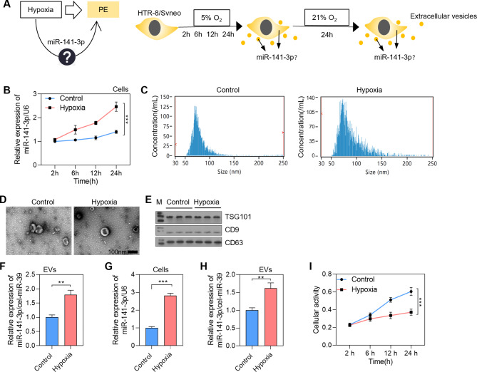
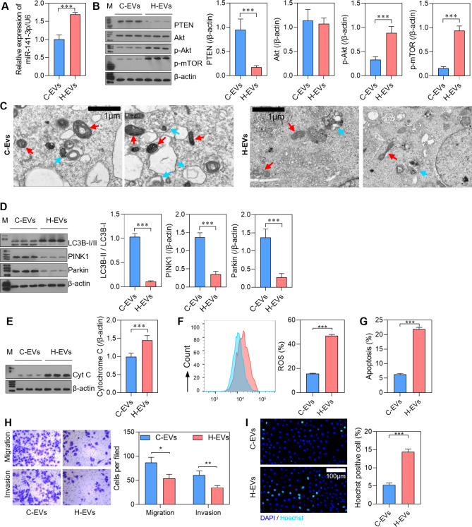
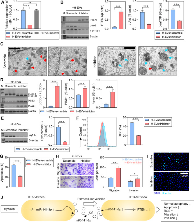
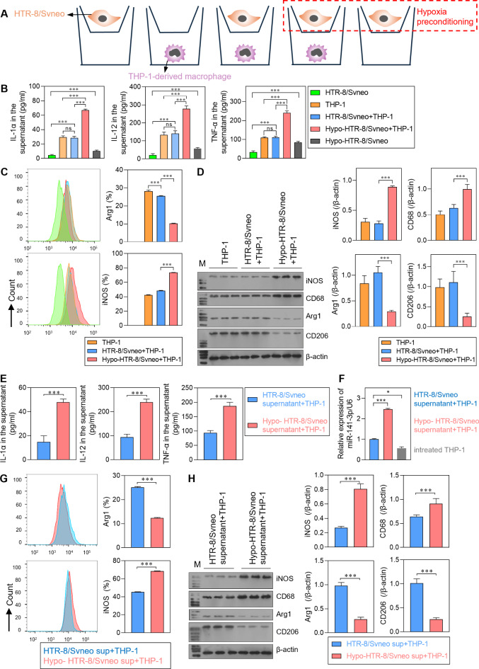
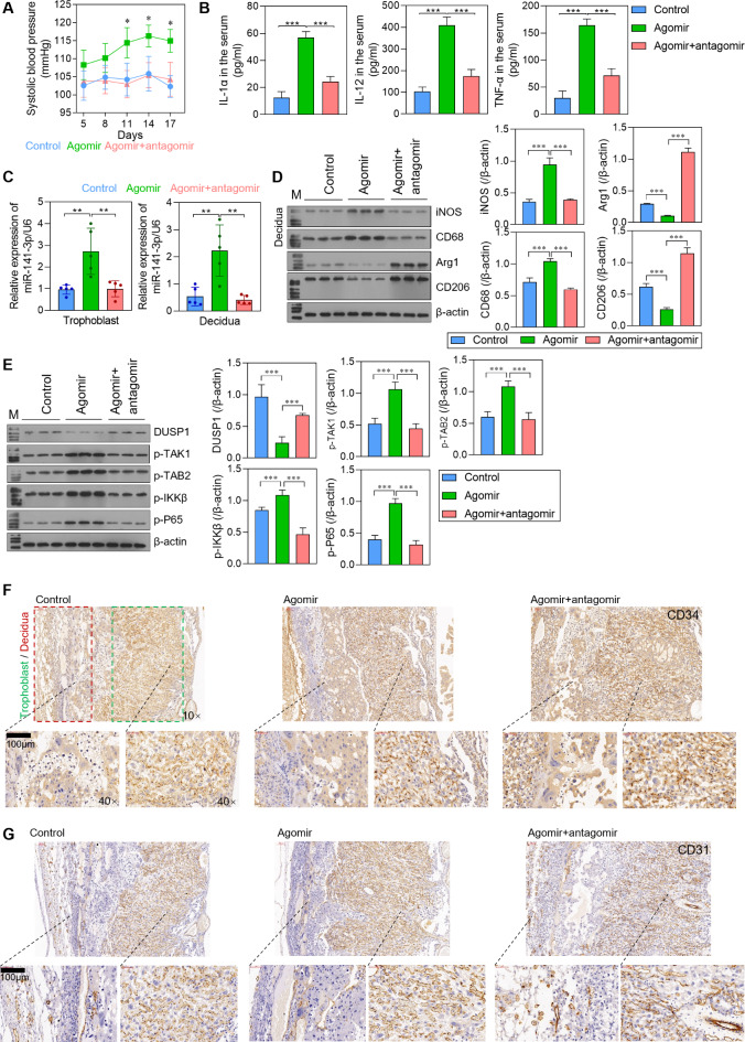
Dysregulation of macrophage polarization can prevent the invasion of trophoblast cells and further limit spiral artery remodeling in preeclampsia (PE). However, its mechanism is obscure. HTR8-/Svneo cells were cultured under normoxic or hypoxic conditions and extracellular vesicles (EVs) in the culture supernatants were extracted. Next, the cells were incubated with those EVs to investigate their effects on trophoblasts. A co-culture system consisting of HTR8-/Svneo cells and macrophages was used to reveal how the trophoblast-derived EVs affected the macrophage subtype. Finally, a PE mouse model and miR-141-3p knockout mice were used to verify the function of miR-141-3p in PE. Hypoxia induced abnormal increases in the levels of miR-141-3p in HTR8-/Svneo cells and EVs. EVs from hypoxia-treated HTR8-/Svneo cells could downregulate PTEN, a potential target of miR-141-3p, and inhibit trophoblast mitophagy and invasion. However, HTR8-/Svneo cells transfected with an miR-141-3p inhibitor could attenuate the influence of EVs. In an HTR8-/Svneo cell plus macrophage co-culture system, hypoxia-pretreated cells promoted the transformation of macrophages into the M1-phenotye, and HTR8-/Svneo invasion was inhibited by the macrophages. MiR-141 from EVs could target and downregulate dual specificity phosphatase 1 (DUSP1) expression in macrophages, induce formation of the M1 macrophage phenotype in THP-1 cells, downregulate DUSP1 expression, and upregulate TAB2/TAK1 signaling. These results were also demonstrated in normal pregnant mice and PE pregnant mice. A hypoxic environment could upregulate miR-141 expression in the EVs of HTR8-/Svneo cells, and THP-1-derived macrophages could uptake EVs releasing miR-141 to downregulate DUSP1 expression and induce the formation of M1 macrophages, which can lead to the development of PE.

Keywords: Angiogenesis; Exosome; Macrophage; Preeclampsia; Trophoblast.


Conflict of interest statement

The authors declare no competing interests.

Figures

Fig. 1
Fig. 1
The levels of miR-141-3p in trophoblast-derived EVs were up-regulated by hypoxia. (A) Schematic diagram showing the simulation of preeclampsia (PE) in vitro. (B) Relative expression of miR-141-3p in HTR-8/Svneo cells cultured under normoxic (Control) and hypoxic conditions for 2, 6, 12, and 24 h. C, D. The morphology of extracted EVs as detected by a NanoSight NS300 device and scanning electron microscope. E. Detection of EVs by western blotting analysis of exosome markers: TSG101, CD9, and CD63. F. HTR-8/Svneo cells were cultured under normoxic and hypoxic conditions for 24 h. Next, the culture medium was replaced and the cells were cultured for another 24 h under normoxic conditions. Finally, the EVs in the culture supernatant were extracted and relative levels of miR-141-3p expression were measured by qRT-PCR. G, H. HTR-8/Svneo cells were cultured under normoxic or hypoxic conditions for 24 h, and then cultured in new medium under normoxic conditions. The relative levels of miR-141-3p expression in cells and EVs were determined by qRT-PCR. I. The viability of HTR-8/Svneo cells cultured under normoxic conditions (Control) and hypoxic conditions for 2, 6, 12, and 24 h as detected by the CCK-8 assay. **p < 0.01; ***p < 0.001.
Fig. 2
Fig. 2
Cellular mitophagy in HTR-8/Svneo cells was inhibited by autocrine EVs. HTR-8/Svneo cells were co-incubated with EVs from HTR-8/Svneo cells cultured under normoxic (C-Exo) or hypoxic (H-Exo) conditions for 24 h. (A) The relative levels of miR-141-3p expression in the C-Exo and H-Exo groups were detected by qRT-PCR. (B) Expression of the potential target of miR-141-3p, PTEN, and the expression of factors in the down-stream pathway. (C) The morphology of cells and autophagosomes as detected by transmission electron microscopy. The red arrow indicates mitochondria, while the yellow arrow indicates mitochondria engulfed by autophagosomes. (D) Detection of autophagy markers, LC3B-I/II, and 2 proteins involved in the mitochondrial autophagy pathway, PINK1 and Parkin, by western blotting. (E) Detection of cytoplasmic Cytochrome C by western blotting. F, G. Detection of ROS levels and apoptotic cells by flow cytometry. H. Cell migration and invasion abilities were detected by Transwell assays (magnification: 200×). I. Cell apoptosis as detected by Hoechst staining. *p < 0.05; **p < 0.01; ***p < 0.001.
Fig. 3
Fig. 3
Cellular mitophagy induced by autocrine EVs in HTR-8/Svneo cells was inhibited by an miR-141-3p inhibitor. HTR-8/Svneo cells were cultured with EVs from hypoxia-pre-treated HTR-8/Svneo cells and transfected with the miR-141-3p scramble (H-Exo + scramble) or inhibitor (H-Exo + inhibitor). (A) Relative expression of miR-141-3p in the H-Exo + scramble and H-Exo + inhibitor groups was detected by qRT-PCR. (B) The expression of PTEN and factors in the down-stream pathway. (C) The morphology of cells and autophagosomes was detected by transmission electron microscopy. The red arrow indicates mitochondria, while the yellow arrow indicates mitochondria engulfed by autophagosomes. (D) Detection of LC3B-I/II, PINK1, and Parkin by western blotting. (E) Detection of cytoplasmic Cytochrome C by western blotting. F, G. Detection of ROS levels and apoptotic cells by flow cytometry. H. Cell migration and invasion abilities were detected by Transwell assays (magnification: 200×). I. Cell apoptosis as detected by Hoechst staining. J. Schematic diagram showing the mechanism of miR-141-3p in HTR-8/Svneo cells. *p < 0.05; **p < 0.01; ***p < 0.001.
Fig. 4
Fig. 4
Effects of co-culture of trophoblasts and macrophages treated with hypoxia on cellular functions. A. THP-1-derived macrophages (lower chamber) were co-cultured with HTR-8/Svneo cells (upper chamber). B. The levels of IL-1α, IL-12, and TNF-α in cell supernatants were detected by ELISA. C. Identification of the macrophage phenotype by flow cytometry. D. Identification of the macrophage phenotype by western blot analysis of phenotype markers: iNOS and CD68 for the M1-phenotype; Arg1 and CD206 for the M2-phenotype. Furthermore, THP-1-derived macrophages were treated with the supernatants of hypoxia-pretreated or non-treated HTR-8/Svneo cells E. The levels of IL-1α, IL-12, and TNF-α in cell supernatants were detected by ELISA. F. Relative expression of miR-141-3p in macrophages. G. Identification of the phenotypes of macrophages by flow cytometry. H. Identification of the phenotypes of macrophages by western blot analysis: iNOS and CD68 for the M1-phenotype; Arg1 and CD206 for the M2-phenotype. HTR-8/Svneo: HTR-8/Svneo cells cultured alone; THP-1: THP-1-derived macrophages cultured alone; HTR-8/Svneo + THP-1: HTR-8/Svneo cells co-cultured with THP-1-derived macrophages; Hyo-HTR-8/Svneo + THP-1: hypoxia-treated HTR-8/Svneo cells co-cultured with THP-1-derived macrophages; HTR-8/Svneo supernatant + THP-1: THP-1-derived macrophages treated with supernatant from HTR-8/Svneo cells; Hypo-HTR-8/Svneo supernatant + THP-1: THP-1-derived macrophages treated with supernatant from hypoxia-treated HTR-8/Svneo cells. ns: no significance; *p < 0.05, ***p < 0.001.
Fig. 5
Fig. 5
MiR-141-3p in HTR-8/Svneo-derived EVs regulated the phenotype of macrophages. THP-1-derived macrophages were stimulated with hypoxia-treated HTR-8/Svneo-derived EVs for 24 h. A. The levels of IL-1α, IL-12, and TNF-α in supernatants were detected by ELISA. B, C. Identification of the phenotypes of macrophages by flow cytometry and western blotting. D. Relative expression of miR-141-3p in macrophages. E. The predicted binding site of miR-141-3p on the DUSP1 3’UTR sequence. F. A dual-luciferase reporter assay was performed to examine the binding site. G-I. DUSP1 expression was detected by qRT-PCR, western blotting, and immunofluorescence. Scramble/ C-Exo: THP-1 derived macrophages transfected with scramble were cultured with EVs from HTR-8/Svneo cells. Scramble/ H-Exo: THP-1 derived macrophages transfected scramble were cultured with EVs from hypoxia-treated HTR-8/Svneo cells. Inhibitor/H-Exo: THP-1 derived macrophages transfected with the miR-141-3p inhibitor were cultured with the supernatants from hypoxia-treated HTR-8/Svneo cells. ns: no significance; ***p < 0.001.
Fig. 6
Fig. 6
The effect of miR-141-3p on macrophages. Macrophages were directly transfected with the miR-141-3p inhibitor or mimics for 24 h. A. The levels of IL-1α, IL-12, and TNF-α in supernatants were detected by ELISA. B, C. Identification of the macrophage phenotype by flow cytometry and western blotting. D. Relative expression of miR-141-3p and DUSP1 in macrophages. E. The expression of DUSP1 and phosphorylated proteins in the down-stream pathway as detected by western blotting. F. Schematic diagram showing the mechanism of miR-141-3p obtained from HTR-8/Svneo-derived EVs in macrophages. ns: no significance; *p < 0.05; ***p < 0.001.
Fig. 7
Fig. 7
The effect of miR-141-3p on regulation of the macrophage phenotype in pregnant mice. (A) Blood pressure was detected on days 5, 8, 11, 14, and 17 of pregnancy using a tail cuff. (B) The levels of IL-1α, IL-12, and TNF-α in serum were detected by ELISA. (C) Relative levels of miR-141-3p expression in trophoblasts and decidua tissues. (D) The phenotypes of macrophages in decidua tissues were detected by western blotting. (E) The expression of DUSP1 and phosphorylated proteins in the down-stream pathway. (F) The placental vasculogenesis markers, CD31 and CD34, as detected by immunohistochemistry. *p < 0.05; **p < 0.01; ***p < 0.001.
Fig. 8
Fig. 8
The effect of miR-141-3p on regulation of the macrophage phenotype in preeclampsia mice. MiR-141-3p-/-(KO) C57BL/6 mice were used for induction of a PE model. (A) Blood pressure was detected on days 5, 8, 11, 14, and 17 of pregnancy using a tail cuff. (B) The levels of IL-1α, IL-12, and TNF-α in serum were detected by ELISA. (C) The phenotypes of macrophages in decidua tissues were detected by western blotting. (D) The expression of DUSP1 and phosphorylated proteins in the down-stream pathway. (E) The placental vasculogenesis markers, CD31 and CD34, as detected by immunohistochemistry. ***p < 0.001.
Fig. 9
Fig. 9
Schematic diagram of the role of miR-141-3p derived from extracellular vesicles in preeclampsia in hypoxic environment.

References

    1. Chappell, L. C., Cluver, C. A., Kingdom, J. & Tong, S. Pre-eclampsia. Lancet. 398 (10297), 341–354 (2021). - PubMed
    1. Ogoyama, M. et al. Non-coding RNAs and prediction of Preeclampsia in the first trimester of pregnancy. Cells. 11, 15 (2022). - PMC - PubMed
    1. Lala, P. K. & Nandi, P. Mechanisms of trophoblast migration, endometrial angiogenesis in preeclampsia: the role of decorin. Cell. Adh Migr. 10(1–2), 111–125 (2016). - PMC - PubMed
    1. Chen, D. B. & Wang, W. Human placental microRNAs and preeclampsia. Biol. Reprod. 88(5), 130 (2013). - PMC - PubMed
    1. Pineles, B. L. et al. Distinct subsets of microRNAs are expressed differentially in the human placentas of patients with preeclampsia. Am. J. Obstet. Gynecol. 196(3), 261e261–261e266 (2007). - PubMed

MeSH terms

LinkOut - more resources