. 2024 Oct 18;14(1):24429.
doi: 10.1038/s41598-024-76196-1.

MicroRNA-130a-3p regulates osimertinib resistance by targeting runt-related transcription factor 3 in lung adenocarcinoma

Affiliations

MicroRNA-130a-3p regulates osimertinib resistance by targeting runt-related transcription factor 3 in lung adenocarcinoma

Takuya Shintani et al. Sci Rep. .

Abstract

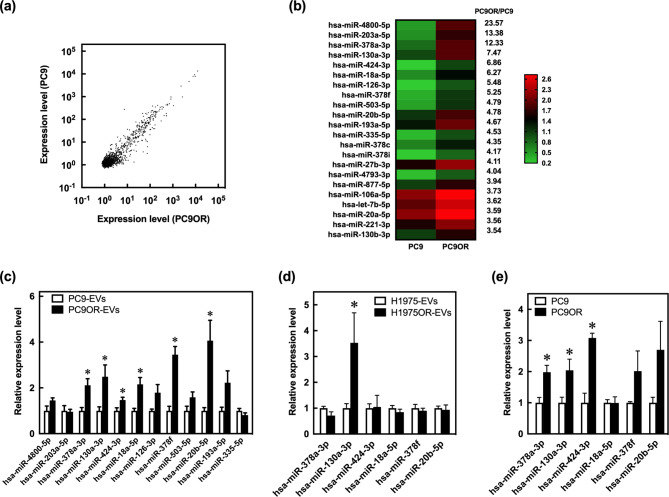
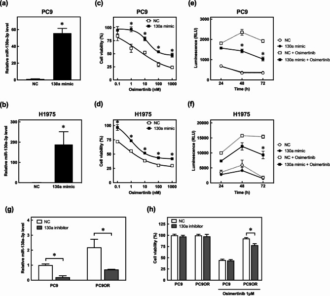
Overcoming resistance to epidermal growth factor receptor tyrosine kinase inhibitors, including osimertinib, is urgent to improve lung cancer treatment outcomes. Extracellular vesicle (EV)-derived microRNAs (EV-miRNAs) play important roles in drug resistance and serve as promising biomarkers. In this study, we aimed to identify EV-miRNAs associated with osimertinib resistance and investigate their clinical relevance. The release of excess EVs was confirmed in the osimertinib-resistant lung adenocarcinoma cell line PC9OR. The exposure of PC9OR-derived EVs and EV-miRNAs to PC9 cells increased cell viability after osimertinib treatment. Microarray analysis revealed that miR-130a-3p was upregulated in EVs derived from PC9OR cells and another osimertinib-resistant cell line (H1975OR). Transfection with miR-130a-3p attenuated osimertinib-induced cytotoxicity and apoptosis in both PC9 and H1975 cells, whereas osimertinib resistance in PC9OR cells was reversed after miR-130a-3p inhibition. Bioinformatics analysis revealed that runt-related transcription factor 3 is a target gene of miR-130a-3p, and it induced osimertinib resistance in PC9 cells. Patients with lower baseline serum miR-130a-3p concentrations had longer progression-free survival. miR-130a-3p is a potential therapeutic target and a predictive biomarker of osimertinib resistance in adenocarcinomas.

Keywords: Extracellular vesicles; Lung adenocarcinoma; MicroRNA-130a-3p; Osimertinib resistance; Runt-related transcription factor 3.


Conflict of interest statement

The authors declare no competing interests.

Figures

Fig. 1
Fig. 1
Characteristics of EVs secreted by PC9 and PC9OR cells in the medium (a) TEM images of isolated EVs from PC9 and PC9OR cells. The black sphere image shows 40-nm gold colloids bound to the CD9 antibody. Scale bars:100 nm. (b) Protein concentrations of CD9 and CD81 (exosomal markers) in isolated EVs from PC9 and PC9OR cells. Original blots are presented in Supplementary Figure S4a. These images show that EVs were isolated (a, b). (c) Size distribution of isolated EVs from PC9 and PC9OR cells. (d) Protein concentrations of isolated total EVs from PC9 and PC9OR cells. (e) Number of CD63- and CD9-positive EVs from PC9 and PC9OR cells. The release of EVs differed in the PC9 and PC9OR cells (c–e). Data are presented as mean ± SEM (n = 3–4) (d, e). *P < 0.05, compared with PC9 cells. P-values were determined using Student’s t-test. EVs, extracellular vesicles; TEM, transmission electron microscopy.
Fig. 2
Fig. 2
Effects of PC9OR-EV- and PC9OR-EV-derived miRNAs on osimertinib resistance in PC9 cells (a) Fluorescence images of PC9 cells after exposure to red-labeled EVs from PC9 (PC9-EVs) and PC9OR cells (PC9OR-EVs). Scale bars: 50 μm. (b) Viability of PC9 cells treated with 5 µg of PC9-EVs and PC9OR-EVs in the absence or presence of 1 µM of osimertinib for 72 h. The controls are PC9 cells treated with PBS (vehicle). (c) The total concentration of miRNA derived from PC9-EVs and PC9OR-EVs. (d) Viability of PC9 cells transfected with 6–12 nM of miRNAs derived from PC9OR-EVs in the absence or presence of 1 µM osimertinib for 72 h. Data are presented as mean ± SEM (n = 3–6) (b–d). *P < 0.05, compared with control or PC9 cells. P-values were determined using Student’s t-test (c) or ANOVA with Tukey–Kramer (b, d). ns, no significant difference. EVs, extracellular vesicles; ANOVA, analysis of variance.
Fig. 3
Fig. 3
Identification of EV-miRNAs associated with osimertinib resistance in lung adenocarcinoma cell lines (a) Scatter plot for the miRNA array analysis of isolated EVs from PC9 and PC9OR cells. (b) A heat map illustrating the expression of 22 EV-miRNAs and demonstrating a 3.5-fold change between PC9 and PC9OR cells. (c) Relative EV-miRNA expression levels in PC9 and PC9OR cells. (d) Relative EV-miRNA expression levels in H1975 and H1975OR cells. (e) Relative intracellular miRNA levels in PC9 and PC9OR cells. miRNA levels were normalized using U6 levels determined by RT-qPCR (c–e). Data are presented as mean ± SEM (n = 5–6). *P < 0.05, compared with PC9 or H1975 cells. P-values were determined using Student’s t-test. EVs, extracellular vesicles; RT-qPCR, reverse transcription-quantitative polymerase chain reaction.
Fig. 4
Fig. 4
Effect of miR-130a-3p on osimertinib resistance in lung adenocarcinoma cell lines (a, b) Relative miR-130a-3p levels in PC9 and H1975 cells transfected with a negative control (NC) and an miR-130a-3p mimic (130a mimic). The miR-130a-3p concentrations were normalized using U6 levels relative to the levels in the cells transfected with the NC. (c, d) Viability of PC9 and H1975 cells transfected with the NC and miR-130a-3p mimic after exposure to various concentrations of osimertinib for 72 h. (e, f) Apoptotic rate in PC9 and H1975 cells after transfection with the NC and miR-130a-3p mimic at 24, 48, and 72 h in the absence or presence of 1 µM osimertinib. (g) Relative miR-130a-3p concentrations in PC9 and PC9OR cells transfected with the NC and miR-130a-3p inhibitor (130a inhibitor). The miR-130a-3p concentrations were normalized using U6 levels relative to the levels in PC9 cells transfected with the NC. (h) Cell viability in PC9 and PC9OR cells transfected with the NC and 130a inhibitor in the absence or presence of 1 µM osimertinib for 72 h. Cell viability is the percentage of viable cells relative to PC9 cells transfected with the NC in the absence of osimertinib. When the standard errors of the means are small, they are contained within the symbols. Data are presented as mean ± SEM (n = 3–12). *P < 0.05, compared with NC. P-values were determined using Student’s t-test (a, b) or ANOVA with Tukey–Kramer (c–h). RLUs, relative luminescence units; ANOVA, analysis of variance.
Fig. 5
Fig. 5
Effect of RUNX3 on osimertinib resistance in PC9 cells (a) Identification of the target genes of miR-130a-3p using miRDB, TargetScan, and miRTarBase databases. (b, c) Protein concentrations of RUNX3 in PC9 and PC9OR cells normalized with β-actin. (d) Protein concentrations of RUNX3 and β-actin in PC9 cells transfected with the negative control (NC) and the miR-130a-3p mimic (130a mimic). (e) Relative mRNA expressions of RUNX3 in PC9 cells transfected with NC and miR-130a-3p inhibitor (130a inhibitor). (f) Protein concentrations of RUNX3 and β-actin in PC9 and PC9OR cells transfected with the NC and the 130a inhibitor. (g) Relative mRNA expression of RUNX3 in PC9 cells transfected with NC and si-RUNX3. The RUNX3 mRNA levels were normalized using the glyceraldehyde-3-phosphate dehydrogenase concentrations relative to the levels in PC9 cells transfected with the NC (e, g). (h) Protein concentrations of RUNX3 and β-actin in PC9 cells transfected with si-RUNX3. (i) Viability of PC9 cells transfected with si-RUNX3 in the absence or presence of 1 µM osimertinib for 72 h. Data are presented as mean ± SEM (n = 3–6). *P < 0.05, compared with PC9 cells or NC. P-values were determined using Student’s t-test (c, e, g) or ANOVA with Tukey–Kramer (i). Original blots are presented in Supplementary Figures S4b-d. ANOVA, analysis of variance.
Fig. 6
Fig. 6
Relationship between serum miR-130a-3p concentration and progression-free survival of patients with NSCLC treated with osimertinib (a) Relative serum miR-130a-3p concentrations in patients with NSCLC. The miR-130a-3p expression indicates the values calculated using the 2−ΔΔCT method and normalized using U6 levels. (b) Progression-free survival of patients with NSCLC treated with first-line osimertinib. Arrows indicate ongoing treatment. * transfer to different institution; # death; AE, discontinuation of treatment due to adverse event; PD, progressive disease; NSCLC, non-small-cell lung cancer.

References

    1. Torre, L. A. et al. Global cancer statistics, 2012. CA Cancer J. Clin.65, 87–108 (2015). - PubMed
    1. Sung, H. et al. Global cancer statistics 2020: GLOBOCAN estimates of incidence and mortality worldwide for 36 cancers in 185 countries. CA Cancer J. Clin.71, 209–249 (2021). - PubMed
    1. Hirsch, F. R. et al. Lung cancer: current therapies and new targeted treatments. Lancet. 389, 299–311 (2017). - PubMed
    1. Thai, A. A., Solomon, B. J., Sequist, L. V., Gainor, J. F. & Heist, R. S. Lung cancer. Lancet. 398, 535–554 (2021). - PubMed
    1. Zhou, W. & Christiani, D. C. East meets West: ethnic differences in epidemiology and clinical behaviors of lung cancer between East asians and caucasians. Chin. J. Cancer. 30, 287–292 (2011). - PMC - PubMed

MeSH terms