. 2024 Oct 19;25(1):980.
doi: 10.1186/s12864-024-10902-1.

Optical genome mapping of structural variants in Parkinson's disease-related induced pluripotent stem cells

Affiliations

Optical genome mapping of structural variants in Parkinson's disease-related induced pluripotent stem cells

Joanne Trinh et al. BMC Genomics. .

Abstract

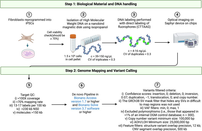
Background: Certain structural variants (SVs) including large-scale genetic copy number variants, as well as copy number-neutral inversions and translocations may not all be resolved by chromosome karyotype studies. The identification of genetic risk factors for Parkinson's disease (PD) has been primarily focused on the gene-disruptive single nucleotide variants. In contrast, larger SVs, which may significantly influence human phenotypes, have been largely underexplored. Optical genomic mapping (OGM) represents a novel approach that offers greater sensitivity and resolution for detecting SVs. In this study, we used induced pluripotent stem cell (iPSC) lines of patients with PD-linked SNCA and PRKN variants as a proof of concept to (i) show the detection of pathogenic SVs in PD with OGM and (ii) provide a comprehensive screening of genetic abnormalities in iPSCs.

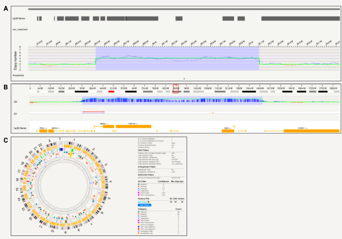
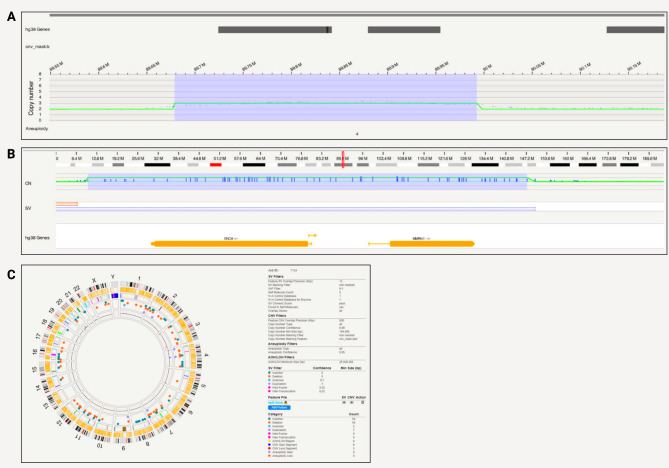
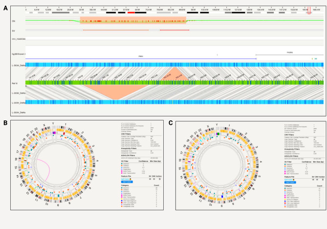
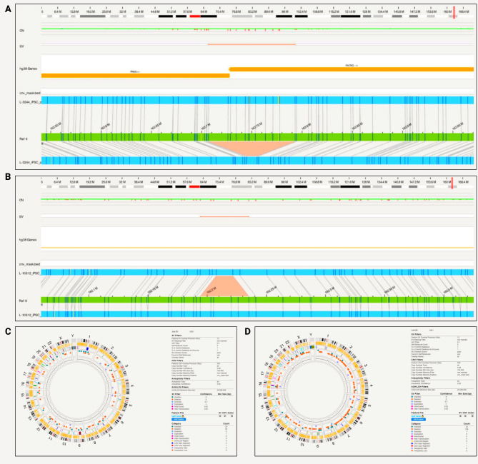
Results: OGM detected SNCA gene triplication and duplication in patient-derived iPSC lines, which were not identified by long-read sequencing. Additionally, various exon deletions were confirmed by OGM in the PRKN gene of iPSCs, of which exon 3-5 and exon 2 deletions were unable to phase with conventional multiplex-ligation-dependent probe amplification. In terms of chromosomal abnormalities in iPSCs, no gene fusions, no aneuploidy but two balanced inter-chromosomal translocations were detected in one line that were absent in the parental fibroblasts and not identified by routine single nucleotide variant karyotyping.

Conclusions: In summary, OGM can detect pathogenic SVs in PD-linked genes as well as reveal genomic abnormalities for iPSCs that were not identified by other techniques, which is supportive for OGM's future use in gene discovery and iPSC line screening.

Keywords: Optical genome mapping; Parkinson’s disease; Structural variants; iPSCs.


Conflict of interest statement

CK serves as a medical advisor to Centogene, Takeda, Retromer Therapeutics, and Lundbeck and received speakers’ honoraria from Bial and Desitin. The remaining authors declare that they have no competing interests.

Figures

Fig. 1
Fig. 1
Overview of Bionano Optical Genomic Mapping Workflow for induced pluripotent stem cells (iPSCs). CV, coefficient of variation; QC, quality check; CNV, copy number variant; kb kilobase; SV, structural variant; VAF, variant allele frequency; AOH/LOH, absence or loss of heterozygosity. Created with BioRender.com
Fig. 2
Fig. 2
SNCA triplication detected with Bionano in a patient-derived iPSC line. A) A triplication spanning 1,696,488 bp that encompasses SNCA; B) Location of the triplication on chromosome 4 at 88,407,893 − 90,104,381 (hg38) that includes genes HERC6, HERC5, PIGY, PYURF, PIGY-DT, HERC3, NAP1L5, FAM13A-AS1, FAM13A, TIGD2, GPRIN3, SNCA, SNCA-AS1, MMRN1; C) Circos plot showing structural variants detected after filtering
Fig. 3
Fig. 3
SNCA duplication detected with Bionano in a patient-derived iPSC line. A) A 313,859 bp long duplication that encompasses SNCA, spanning positions 89,678,642 − 89,992,501 on chromosome 4; B) Location of the triplication on chromosome 4 at 85388502–89998264 (hg38) that includes genes SNCA, SNCA-AS1, MMRN1; C) Circos plot showing structural variants detected after filtering
Fig. 4
Fig. 4
PRKN compound heterozygous variants detected with Bionano in iPSCs and the parental fibroblast. A) The compound heterozygous exon 2 and exon 3–5 deletion was captured with phase. The PRKN exon 2 deletion starts at position 162,338,358 and ends at 162,450,583, the PRKN exon 3–5 deletion starts at 162,029,361 and ends at 162,279,161 and partially overlaps with PACRG; B) Circos plot showing a translocation and other structural variants detected after filtering in the iPSC line from patient iPS-L-3034; C) Circos plot without the translocation present and structural variants detected after filtering in the parental fibroblast line L-3034
Fig. 5
Fig. 5
PRKN compound heterozygous variants detected with Bionano in iPSCs. A) A patient-derived iPSC line (iPS-L-3244) with PRKN exon 1 deletion. PRKN exon 1 deletion starts at position 162,716,506 and ends at 162,792,085 and partially overlaps with PACRG.; B) A patient-derived iPSC line (iPS-L-10312) with PRKN exon 4 deletion. PRKN exon 4 deletion starts at position 162,198,660 and ends at 162,225,645; C) Circos plot showing structural variants detected after filtering in iPS-L-3244; D) Circos plot with structural variants detected after filtering in iPS-L-10312

References

    1. Ho SS, Urban AE, Mills RE. Structural variation in the sequencing era. Nat Rev Genet. 2020;21(3):171–89. - PMC - PubMed
    1. Skrahina V, Gaber H, Vollstedt EJ, Forster TM, Usnich T, Curado F, et al. The Rostock International Parkinson’s Disease (ROPAD) study: protocol and initial findings. Mov Disord. 2021;36(4):1005–10. - PMC - PubMed
    1. Nalls MA, Blauwendraat C, Vallerga CL, Heilbron K, Bandres-Ciga S, Chang D, et al. Identification of novel risk loci, causal insights, and heritable risk for Parkinson’s disease: a meta-analysis of genome-wide association studies. Lancet Neurol. 2019;18(12):1091–102. - PMC - PubMed
    1. Farrer M, Maraganore DM, Lockhart P, Singleton A, Lesnick TG, de Andrade M, et al. Alpha-synuclein gene haplotypes are associated with Parkinson’s disease. Hum Mol Genet. 2001;10(17):1847–51. - PubMed
    1. Singleton AB, Farrer M, Johnson J, Singleton A, Hague S, Kachergus J, et al. Alpha-synuclein locus triplication causes Parkinson’s disease. Science. 2003;302(5646):841. - PubMed

LinkOut - more resources