. 2024 Oct 18;21(1):263.
doi: 10.1186/s12974-024-03256-8.

Intestinal homeostasis disrupted by Periodontitis exacerbates Alzheimer's Disease in APP/PS1 mice

Affiliations

Intestinal homeostasis disrupted by Periodontitis exacerbates Alzheimer's Disease in APP/PS1 mice

Xueshen Qian et al. J Neuroinflammation. .

Abstract

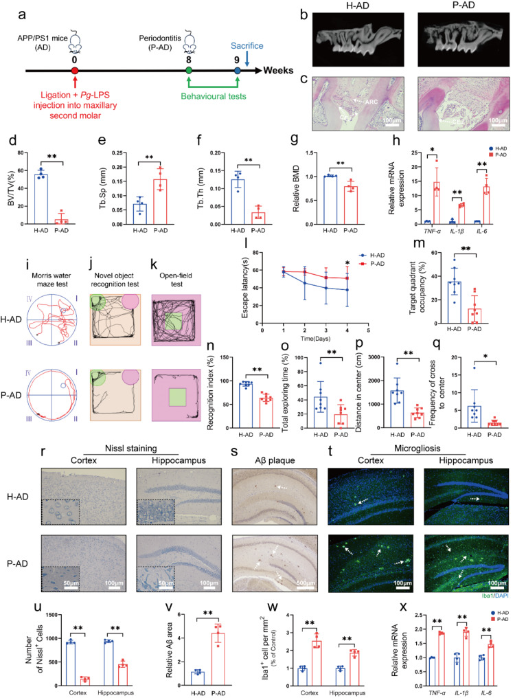
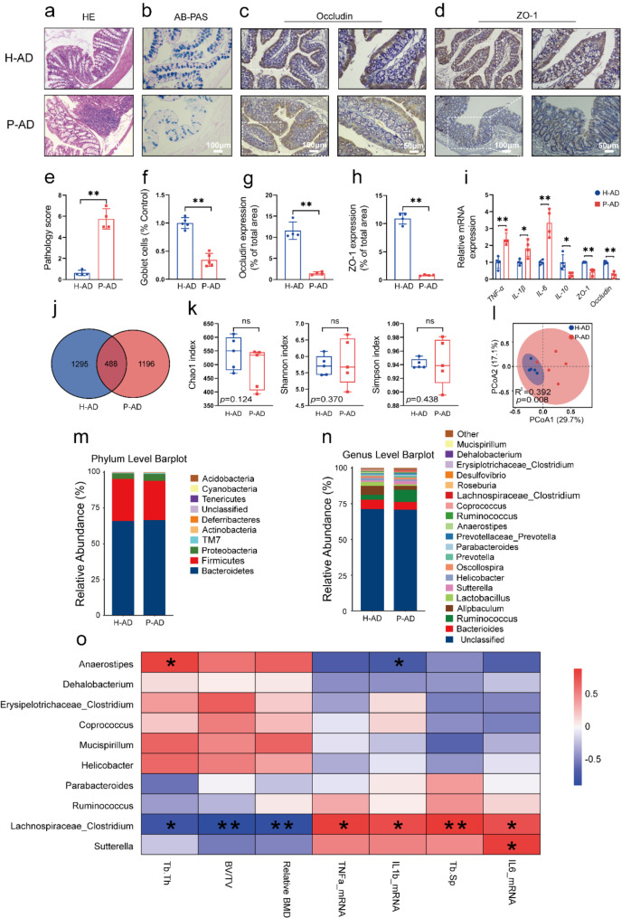
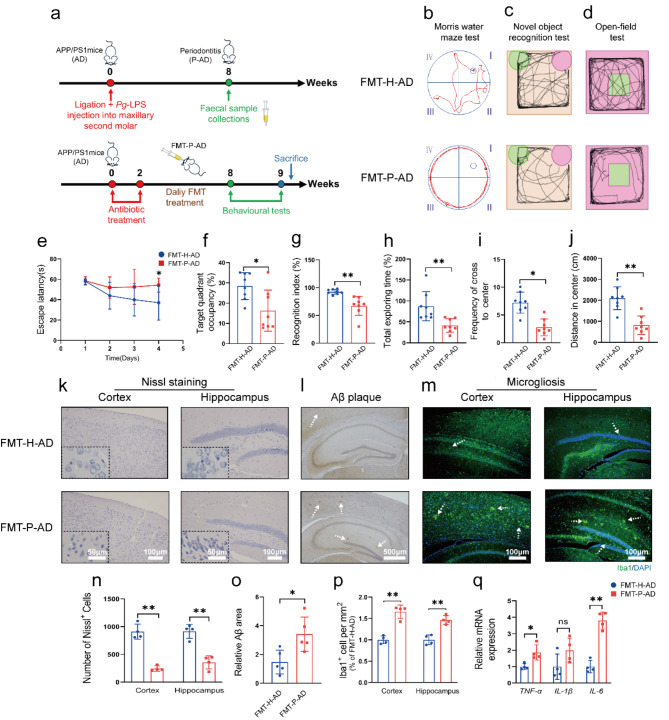
Periodontitis exacerbates Alzheimer's disease (AD) through multiple pathways. Both periodontitis and AD are intricately correlated to intestinal homeostasis, yet there is still a lack of direct evidence regarding whether periodontitis can regulate the progression of AD by modulating intestinal homeostasis. The current study induced experimental periodontitis in AD mice by bilaterally ligating the maxillary second molars with silk and administering Pg-LPS injections in APPswe/PS1ΔE9 (APP/PS1) mice. Behavioral tests and histological analyses of brain tissue were conducted after 8 weeks. Gut microbiota was analyzed and colon tissue were also evaluated. Then, fecal microbiota from mice with periodontitis was transplanted into antibiotic-treated mice to confirm the effects of periodontitis on AD and the potential mechanism was explored. The results indicated periodontitis exacerbated cognitive impairment and anxious behaviour in APP/PS1 mice, with increased Aβ deposition, microglial overactivation and neuroinflammation in brain. Moreover, the intestinal homeostasis of AD mice was altered by periodontitis, including affecting gut microbiota composition, causing colon inflammation and destroyed intestinal epithelial barrier. Furthermore, AD mice that underwent fecal transplantation from mice with periodontitis exhibited worsened AD progression and disrupted intestinal homeostasis. It also impaired intestinal barrier function, elevated peripheral inflammation, damaged blood-brain barrier (BBB) and caused neuroinflammation and synapses impairment. Taken together, the current study demonstrated that periodontitis could disrupt intestinal homeostasis to exacerbate AD progression potential via causing gut microbial dysbiosis, intestinal inflammation and intestinal barrier impairment to induce peripheral inflammation and damage BBB, ultimately leading to neuroinflammation and synapse impairment. It underscores the importance of maintaining both periodontal health and intestinal homeostasis to reduce the risk of AD.

Keywords: Alzheimer’s disease; Gut microbiota; Inflammation; Intestinal homeostasis; Periodontitis.


Conflict of interest statement

The authors declare no competing interests.

The authors assert that they possess no conflicts of interest.

Figures

Fig. 1
Fig. 1
Periodontitis exacerbated behavioral disorders and AD-like pathological lesions in the APP/PS1 mice. (a) Design of the experiment. (b) Mesial-distal micro-CT slices of maxillary molars. (c) HE staining of the alveolar bone. (d-g) Quantitative analysis of micro-CT data. (h) The mRNA levels of TNF-α, IL-1β and IL-6 in gingiva. (i) Diagrams showing the swimming paths during the spatial probe period. (j) Mice exploring new objects and familiar object path maps. (k) The open-field tests included representative tracking images of movement. (l) Escape latency of mice to reach the hidden platform. (m) Percentage of time utilized by the mice for swimming in the target quadrant. (n) New object RI. (o) Total time spent exploring new objects. (p) Distance of the mice in central area. (q) Frequency of the mice of the cross to center. (r) Representative images of Nissl staining. (s) Representative images of Aβ immunostaining. (t) Representative images of Iba1 immunostaining. (u) Quantitative analysis of the number of surviving neurons in cortex and hippocampus. (v) Area fractions of relative Aβ-positive in hippocampus. (w) Iba1-positive area fractions in cortex and hippocampus. (x) The relative mRNA expression levels of TNF-α, IL-1β and IL-6 in Cortex. ∗ p < 0.05, ∗∗ p < 0.01; ARC: alveolar ridge crest; CEJ: cementoenamel junction
Fig. 2
Fig. 2
Periodontitis led to disruptions of intestinal homeostasis in the APP/PS1 mice. (a) HE staining of colon. (b) AB-PAS staining. (c) Representative images of ZO-1 immunostaining. (d) Representative images of Occludin immunostaining. (e) Histological score of the colon. (f) Quantitative analysis of Goblet cells. (g-h) Quantitative analysis of ZO-1 and Occludin expression. (i) The relative mRNA expression levels of TNF-α, IL-6, IL-1β, IL-10, ZO-1 and Occludin in the colon. (j) Venn diagram showing the difference in ASV between the H-AD and P-AD groups. (k) The α diversity of Chao1 index, Shannon index and Simpson index. (l) PCoA analysis. (m-n) Periodontitis changes the overall composition of the gut microbiota in phylum (m) and genus levels (n). (o) Spearman correlation analysis between gut microbiota and parameters related to periodontitis. ∗ p < 0.05, ∗∗ p < 0.01, ns: not significant
Fig. 3
Fig. 3
Transplantation of periodontitis-altered gut microbiota worsened behavioral disorders and AD-like pathology in AD mice. (a) Design of the experiment. (b) Diagrams showing the swimming paths during the spatial probe period. (c) Mice explore new objects and familiar object path maps. (d) The open-field test included representative tracking images of movement. (e) Escape latency of mice to reach the hidden platform. (f) Percentage of time utilized by the mice for swimming in the target quadrant. (g) New object RI. (h) Total time spent exploring new objects. (i) Frequency of the number of times the mice cross to center. (j) Distance of the mice in central area. (k) Representative images of Nissl staining. (l) Representative images of Aβ immunostaining. (m) Representative images of Iba1 immunofluorescence staining. (n) Quantitative analysis of the number of surviving neurons in cortex and hippocampus. (o) Fractions of Aβ-positive area. (p) Quantitative analysis of the area fraction in the cortex and hippocampus. (q) The relative mRNA expression levels of TNF-α, IL-1β and IL-6 in the Cortex. ∗ p < 0.05, ∗∗ p < 0.01
Fig. 4
Fig. 4
Transplantation of periodontitis-altered gut microbiota into AD mice caused disruptions of intestinal homeostasis. (a) HE staining of colon. (b) AB-PAS staining. (c) Representative images of ZO-1 immunostaining. (d) Representative images of Occludin immunostaining. (e) Histologic score of the colon. (f) Quantitative analysis of Goblet cells. (f) Quantitative analysis of Goblet cells. (g-h) Quantitative analysis of ZO-1 and Occludin expression. (i) The relative mRNA expression of TNF-α, IL-1β, IL-6, IL-10, ZO-1 and Occludin in the colon. (j) Venn diagram showing the difference in ASV between the FMT-H-AD and FMT-P-AD groups. (k) The α diversity of Chao1 index, Shannon index and Simpson index. (l) PCoA analysis. (m-n) Transplantation of periodontitis altered the overall composition of the gut microbiota at phylum (m) and genus (n) levels). ∗ p < 0.05, ∗∗ p < 0.01 versus the FMT-H-AD group
Fig. 5
Fig. 5
The potential mechanisms of disruptions of intestinal homeostasis on exacerbation of AD by periodontitis. (a) Representative images of F4/80 immunostaining. (b) Quantification of F4/80-positive area fractions in the colon. (c-d) The levels of LPS, TNF-α and IL-1β in colon. (e-f) The levels of LPS, TNF-α and IL-1β in peripheral blood. (g) Representative images of TEM with BBB. The arrow shows the astrocytic end-feet swelling. (h-i) Western blot and quantitative analysis of ZO-1 and Occludin in Cortex. (j-k) The levels of LPS, TNF-α and IL-1β in hippocampus. (l) Representative images of TEM with synapses. The arrow shows blurring of synaptic cleft structure. (m-n) Immunofluorescence localization and quantification of Iba1 (green) and Aβ (magenta) in cortex and hippocampus. (o) Spearman correlation analysis with intestinal lesions, brain AD-like lesions, peripheral blood inflammation and gut microbiota. ∗ p < 0.05, ∗∗ p < 0.01
Scheme 1
Scheme 1
A schematic illustrates how periodontitis exacerbated AD progression potential via causing gut microbial dysbiosis, intestinal inflammation and intestinal barrier impairment to induce peripheral inflammation and damage BBB, ultimately causing neuroinflammation and synapse impairment

References

    1. Slots J. Periodontitis: facts, fallacies and the future. Periodontol 2000. 2017;75:7–23. - PubMed
    1. Jepsen S, Suvan J, Deschner J. The association of periodontal diseases with metabolic syndrome and obesity. Periodontol 2000. 2020;83:125–53. - PubMed
    1. Sanz M, Marco Del Castillo A, Jepsen S, Gonzalez-Juanatey JR, D’Aiuto F, Bouchard P, et al. Periodontitis and cardiovascular diseases: Consensus report. J Clin Periodontol. 2020;47:268–88. - PMC - PubMed
    1. Lalla E, Papapanou PN. Diabetes mellitus and periodontitis: a tale of two common interrelated diseases. Nat Rev Endocrinol. 2011;7:738–48. - PubMed
    1. Carballo Á, López-Dequidt I, Custodia A, Botelho J, Aramburu-Núñez M, Machado V, et al. Association of periodontitis with cognitive decline and its progression: contribution of blood-based biomarkers of Alzheimer’s disease to this relationship. J Clin Periodontol. 2023;50:1444–54. - PubMed

Substances

LinkOut - more resources