Review
. 2024 Dec;12(6):e1219.
doi: 10.1002/prp2.1219.

Therapeutic potential of agents targeting cannabinoid type 2 receptors in organ fibrosis

Affiliations
Review

Therapeutic potential of agents targeting cannabinoid type 2 receptors in organ fibrosis

Lujain Bader Eddin et al. Pharmacol Res Perspect. 2024 Dec.

Abstract

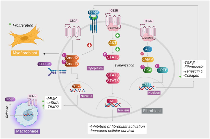
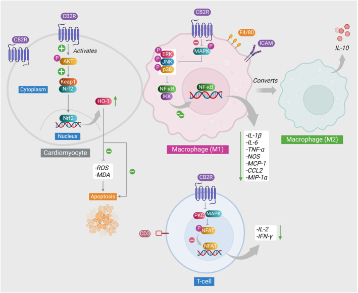
The endocannabinoid system has garnered attention as a potential therapeutic target in a range of pathological disorders. Cannabinoid receptors type 2 (CB2) are a class of G protein-coupled receptors responsible for transmitting intracellular signals triggered by both endogenous and exogenous cannabinoids, including those derived from plants (phytocannabinoids) or manufactured synthetically (synthetic cannabinoids). Recent recognition of the role of CB2 receptors in fibrosis has fueled interest in therapeutic targeting of CB2 receptors in fibrosis. Fibrosis is characterized by the alteration of the typical cellular composition within the tissue parenchyma, resulting from exposure to diverse etiological factors. The pivotal function of CB2 agonists has been widely recognized in the regulation of inflammation, fibrogenesis, and various other biological pathologies. The modulation of CB2 receptors, whether by enhancing their expression or activating their function, has the potential to provide benefits in numerous conditions, particularly by avoiding any associated adverse effects on the central nervous system. The sufficient activation of CB2 receptors resulted in the complete suppression of gene expression related to transforming growth factor β1 and its subsequent fibrogenic response. Multiple reports have also indicated the diverse functions that CB2 agonists possess in mitigating chronic inflammation and subsequent fibrosis development in various types of tissues. While currently in the preclinical stage, the advancement of CB2 compounds has garnered significant attention within the realm of drug discovery. This review presents a comprehensive synthesis of various independent experimental studies elucidating the pivotal role of identified natural and synthetic CB2 agonists in the pathophysiology of organ fibrosis, specifically in the cardiac, hepatic, and renal systems.

Keywords: CB2 agonists; cannabinoids; cardiac fibrosis; fibrosis; phytocannabinoids; renal fibrosis.


Conflict of interest statement

The authors declare no conflict of interest.

Figures

FIGURE 1
FIGURE 1
The protective effects of CB2 receptors on fibroblasts. Upon the activation of CB2 receptors following the binding of CB2 agonists, several intracellular pathways can be initiated. As CB2 receptor is a GPCR bound to Gi subunit this leads to the inhibition of AC that in turn leads to a decrease in the intracellular concentration of cyclic adenosine monophosphate (cAMP). Decreased cAMP inhibited the activation and phosphorylation of PKA which in turn inhibits the phosphorylation and nuclear translocation of CREB transcription factor which ultimately inhibits the gene expression of fibrogenic proteins including tumor growth factor‐beta (TGF‐β), fibronectin, collagen, and tenascin C. The decreased production of TGF‐β results in decreased activation of TGF‐βR that positively acts on transducing Smad2 and Smad3 signaling and producing TIMP2 and α‐SMA, and decreased matrix metalloproteinase release. CB2 receptor also directly inhibit the action of Smad2 and Smad3 transcription factors. Besides, CB2 receptor activation increases activation of AKT, which in turn phosphorylates STAT3 transcription factor. Upon phosphorylation, STAT3 induces transcription of protective genes enhancing cellular survival. A side illustration of CB2 receptor expression on macrophages and the ultimate effect of inhibited PDGF production following CB2 receptor activation. AC, adenylyl cyclase; AKT, protein kinase B; cAMP, adenosine 3′;5′‐cyclic monophosphate; CREB, cAMP response element‐binding protein; PDGF, platelet‐derived growth factor; PKA, protein kinase A; Smad, suppressor of mothers against decapentaplegic; STAT3, signal transducer and activator of transcription 3.
FIGURE 2
FIGURE 2
CB2 receptors' mechanisms of action in cardiomyocytes and inflammatory cells. As illustrated, the activation of CB2 receptors in cardiomyocytes phosphorylates AKT which degrades Keap1 leading to the dissociation of Nrf2 and its translocation to the nucleus where it increases the transcription of HO‐1 gene. This decreases the generated ROS, and peroxidized lipids and inhibits apoptosis. In the macrophage, CB2 activation suppresses the activation of mitogen‐activated protein kinases (MAPK) and its downstream mediators. Consequently, NF‐κB is inhibited and there will be no dissociation from its inhibitor complex with IKK, and the release of inflammatory mediators is inhibited. With an unknown mechanism activated CB2 receptor causes the polarization of M1 macrophages into the anti‐inflammatory phenotype M2 macrophages which contribute to the anti‐inflammatory response through the release of anti‐inflammatory cytokine IL‐10. In T cells, activated CB2 receptor deactivates MAPK pathway, inhibiting PKC. As a result, NFAT phosphorylation, activation, and nuclear translocation are inhibited, resulting in a reduced release of IL‐2 and INF‐γ. AKT, protein kinase B; CCL2, chemokine (C‐C motif) ligand 2; ERK, extracellular signal‐regulated kinase; HO‐1, heme oxygenase 1; IKK, inhibitor of nuclear factor‐κB (IκB) kinase; IL‐10, interleukin‐10; IL‐1β, interleukin‐1β; IL‐6, interleukin‐6; IL‐2, interleukin‐2; INF‐γ, interferon gamma; JNK, c‐Jun N‐terminal kinase; keap1, Kelch‐like ECH‐associated protein 1; MAPK, mitogen‐activated protein kinase; MCP‐1, monocyte chemoattractant protein‐1; MDA, malondialdehyde; MIP‐1α, macrophage inflammatory protein‐1 alpha; NF‐κB, nuclear factor kappa‐light‐chain enhancer of activated B cells; NOS, nitric oxide synthase; Nrf2, nuclear factor erythroid 2‐related factor 2; ROS, reactive oxygen species; TNF‐α, tumor necrosis factor alpha.

References

    1. Horowitz JC, Thannickal VJ. Mechanisms for the resolution of organ fibrosis. Phys Ther. 2018;34:43‐55. doi:10.1152/physiol.00033.2018 - DOI - PMC - PubMed
    1. Galliot B, Crescenzi M, Jacinto A, Tajbakhsh S. Trends in tissue repair and regeneration. Development. 2017;144:357‐364. doi:10.1242/dev.144279 - DOI - PubMed
    1. Wynn TA. Cellular and molecular mechanisms of fibrosis. J Pathol. 2008;214:199‐210. doi:10.1002/path.2277 - DOI - PMC - PubMed
    1. Suthahar N, Meijers WC, Silljé HHW, de Boer RA. From inflammation to fibrosis‐molecular and cellular mechanisms of myocardial tissue Remodelling and perspectives on differential treatment opportunities. Curr Heart Fail Rep. 2017;14:235‐250. doi:10.1007/s11897-017-0343-y - DOI - PMC - PubMed
    1. Fonseca BM, Costa MA, Almada M, Correia‐da‐Silva G, Teixeira NA. Endogenous cannabinoids revisited: a biochemistry perspective. Prostaglandins Other Lipid Mediat. 2013;102‐103:13‐30. doi:10.1016/j.prostaglandins.2013.02.002 - DOI - PubMed

MeSH terms

LinkOut - more resources