. 2025 Mar;35(3):1242-1254.
doi: 10.1007/s00330-024-11140-5. Epub 2024 Oct 19.

Preoperative prediction of diffuse glioma type and grade in adults: a gadolinium-free MRI-based decision tree

Affiliations

Preoperative prediction of diffuse glioma type and grade in adults: a gadolinium-free MRI-based decision tree

Aynur Azizova et al. Eur Radiol. 2025 Mar.

Abstract

Objectives: To develop a gadolinium-free MRI-based diagnosis prediction decision tree (DPDT) for adult-type diffuse gliomas and to assess the added value of gadolinium-based contrast agent (GBCA) enhanced images.

Materials and methods: This study included preoperative grade 2-4 adult-type diffuse gliomas (World Health Organization 2021) scanned between 2010 and 2021. The DPDT, incorporating eleven GBCA-free MRI features, was developed using 18% of the dataset based on consensus readings. Diagnosis predictions involved grade (grade 2 vs. grade 3/4) and molecular status (isocitrate dehydrogenase (IDH) and 1p/19q). GBCA-free diagnosis was predicted using DPDT, while GBCA-enhanced diagnosis included post-contrast images. The accuracy of these predictions was assessed by three raters with varying experience levels in neuroradiology using the test dataset. Agreement analyses were applied to evaluate the prediction performance/reproducibility.

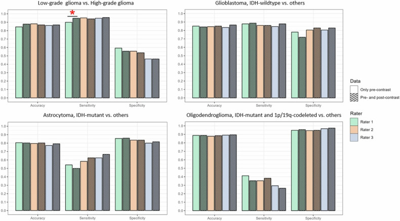
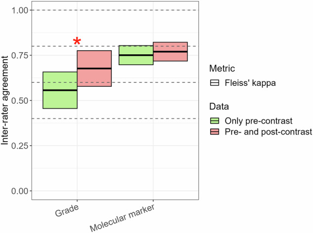
Results: The test dataset included 303 patients (age (SD): 56.7 (14.2) years, female/male: 114/189, low-grade/high-grade: 54/249, IDH-mutant/wildtype: 82/221, 1p/19q-codeleted/intact: 34/269). Per-rater GBCA-free predictions achieved ≥ 0.85 (95%-CI: 0.80-0.88) accuracy for grade and ≥ 0.75 (95%-CI: 0.70-0.80) for molecular status, while GBCA-enhanced predictions reached ≥ 0.87 (95%-CI: 0.82-0.90) and ≥ 0.77 (95%-CI: 0.71-0.81), respectively. No accuracy difference was observed between GBCA-free and GBCA-enhanced predictions. Group inter-rater agreement was moderate for GBCA-free (0.56 (95%-CI: 0.46-0.66)) and substantial for GBCA-enhanced grade prediction (0.68 (95%-CI: 0.58-0.78), p = 0.008), while substantial for both GBCA-free (0.75 (95%-CI: 0.69-0.80) and GBCA-enhanced (0.77 (95%-CI: 0.71-0.82), p = 0.51) molecular status predictions.

Conclusion: The proposed GBCA-free diagnosis prediction decision tree performed well, with GBCA-enhanced images adding little to the preoperative diagnostic accuracy of adult-type diffuse gliomas.

Key points: Question Given health and environmental concerns, is there a gadolinium-free imaging protocol to preoperatively evaluate gliomas comparable to the gadolinium-enhanced standard practice? Findings The proposed gadolinium-free diagnosis prediction decision tree for adult-type diffuse gliomas performed well, and gadolinium-enhanced MRI demonstrated only limited improvement in diagnostic accuracy. Clinical relevance Even inexperienced raters effectively classified adult-type diffuse gliomas using the gadolinium-free diagnosis prediction decision tree, which, until further validation, can be used alongside gadolinium-enhanced images to respect standard practice, despite this study showing that gadolinium-enhanced images hardly improved diagnostic accuracy.

Keywords: Brain neoplasms; Gadolinium; Glioma; Isocitrate dehydrogenase; Magnetic resonance imaging.


Conflict of interest statement

Compliance with ethical standards. Guarantor: The scientific guarantor of this publication is Vera C. Keil. Conflict of interest: The authors of this manuscript declare relationships with the following companies: V.C.K.: Lilly Pharma (Consulting); Junior Deputy Editor for European Radiology, as such they did not participate in the selection or review processes for this article. The remaining authors declare no conflicts of interest. Statistics and biometry: One of the authors has significant statistical expertise. Informed consent: Written informed consent was waived by the Institutional Review Board. Ethical approval: Institutional Review Board approval was obtained. Study subjects or cohorts overlap: The same study cohort was used in another study, which was recently accepted by the Journal of Neuroimaging on August 14, 2024. The in-press study explored a different research question focused on predicting glioma enhancement features using the proposed Enhancement Prediction Decision Tree. Methodology: Retrospective Observational Performed at one institution

Figures

Fig. 1
Fig. 1
Diagnosis prediction decision tree (DPDT) based on GBCA-free MRI sequences. Flow chart describes DPDT for adult-type diffuse gliomas encompassing seven VASARI (necrosis, diffusion, hemorrhage, non-enhancing tumor margin, calvarial remodeling, cysts, proportion of edema) and four non-VASARI (T2-FLAIR mismatch sign, T2 signal homogeneity, calcification, midline shift) imaging features guiding the histomolecular diagnosis decision. GBCA, gadolinium-based contrast agent; VASARI, Visually AcceSAble Rembrandt Images
Fig. 2
Fig. 2
Patient enrollment diagram. The flow chart depicts the patients included and excluded in this study. IDH, isocitrate dehydrogenase
Fig. 3
Fig. 3
Per-rater prediction performance of histomolecular diagnosis of adult-type diffuse gliomas using GBCA-free vs. GBCA-enhanced scans. Color bar charts show the prediction performance, including accuracy, sensitivity, and specificity levels in predicting tumor grade, IDH mutation, and 1p/19q-codeletion status using GBCA-free and GBCA-enhanced scans per rater (rater 1 = green bar, rater 2 = orange bar, rater 3 = blue bar). Comparison of GBCA-free and GBCA-enhanced predictions revealed no significant differences (p > 0.05) except for rater 1’s sensitivity in histopathological grade prediction (red star, p = 0.006). GBCA, gadolinium-based contrast agent
Fig. 4
Fig. 4
Group inter-rater agreement in histomolecular diagnosis prediction of adult-type diffuse gliomas using GBCA-free vs. GBCA-enhanced scans. The color box plot shows inter-rater agreement in predicting tumor grade (low-grade: grade 2 vs. high-grade: grade 3/4) and molecular status (astrocytoma, IDH-mutant vs. oligodendroglioma, IDH-mutant and 1p/19q-codeleted vs. glioblastoma, IDH-wildtype) among all raters. Green bars depict the results based on the evaluation of GBCA-free scans (only pre-contrast sequences: pre-contrast T1-weighted, T2-weighted, FLAIR, DWI/ADC, and SWI (if present)), and red bars show the results of the evaluation using GBCA-enhanced scans (pre- and post-contrast sequences: pre-contrast T1-weighted, T2-weighted, FLAIR, DWI/ADC, SWI (if present) + post-contrast T1-weighted). Comparison of agreements between GBCA-free and GBCA-enhanced predictions was significant for tumor grade (red star, p = 0.008), while it was insignificant for molecular marker (p > 0.05). Note: The interpretation of agreement values was as follows: 0.01–0.20, slight; 0.21–0.40, fair; 0.41–0.60, moderate; 0.61–0.80, substantial; and 0.81–0.99, almost perfect. GBCA, gadolinium-based contrast agent; IDH, isocitrate dehydrogenase
Fig. 5
Fig. 5
Intra-rater inter-group agreement in histomolecular diagnosis prediction of adult-type diffuse gliomas. The color box plot describes intra-rater inter-group agreement comparing GBCA-free and GBCA-enhanced histomolecular diagnosis predictions of each rater (rater 1 = green bar, rater 2 = orange bar, rater 3 = blue bar). Diagnosis predictions include tumor grade (low-grade: grade 2 vs. high-grade: grade 3/4) and molecular status (astrocytoma, IDH-mutant vs. oligodendroglioma, IDH-mutant and 1p/19q-codeleted vs. glioblastoma, IDH-wildtype) evaluations. Red triangles indicate prevalence-adjusted and bias-adjusted kappa (PABAK) values that compensate for the possible influence of dataset diagnosis imbalances. Comparison of agreements among all raters revealed significant differences for both tumor grade and molecular marker (p < 0.001). Note: The interpretation of agreement values was as follows: 0.01–0.20, slight; 0.21–0.40, fair; 0.41–0.60, moderate; 0.61–0.80, substantial; and 0.81–0.99, almost perfect. GBCA, gadolinium-based contrast agent; IDH, isocitrate dehydrogenase
Fig. 6
Fig. 6
Group inter-rater agreement in evaluating imaging features included in the Diagnosis Prediction Decision tree (DPDT) for adult-type diffuse gliomas. The color box plot shows inter-rater agreement in evaluating single DPDT imaging features using GBCA-free or GBCA-enhanced scans among all raters. Green bars depict the results based on the assessment of GBCA-free scans (only pre-contrast sequences: pre-contrast T1-weighted, T2-weighted, FLAIR, DWI/ADC, and SWI (if present)), and red bars show the results of the evaluation using GBCA-enhanced scans (pre- and post-contrast sequences: pre-contrast T1-weighted, T2-weighted, FLAIR, DWI/ADC, SWI (if present) + post-contrast T1-weighted). Comparison of agreements between GBCA-free and GBCA-enhanced assessments was insignificant (p > 0.05) except for hemorrhage and midline shift (red stars, p = 0.02 and p = 0.04, respectively). Note: The interpretation of agreement values was as follows: 0.01–0.20, slight; 0.21–0.40, fair; 0.41–0.60, moderate; 0.61–0.80, substantial; and 0.81–0.99, almost perfect. GBCA, gadolinium-based contrast agent

References

    1. Ellingson BM, Bendszus M, Boxerman J et al (2015) Consensus recommendations for a standardized Brain Tumor Imaging Protocol in clinical trials. Neuro Oncol 17:1188–1198 - PMC - PubMed
    1. Haddad AF, Young JS, Morshed RA, Berger MS (2022) FLAIRectomy: resecting beyond the contrast margin for glioblastoma. Brain Sci 12:544 - PMC - PubMed
    1. Castet F, Alanya E, Vidal N et al (2019) Contrast-enhancement in supratentorial low-grade gliomas: a classic prognostic factor in the molecular age. J Neurooncol 143:515–523 - PubMed
    1. Eichberg DG, Di L, Morell AA et al (2020) Incidence of high grade gliomas presenting as radiographically non-enhancing lesions: experience in 111 surgically treated non-enhancing gliomas with tissue diagnosis. J Neurooncol 147:671–679 - PubMed
    1. Wen PY, van den Bent M, Youssef G et al (2023) RANO 2.0: update to the response assessment in neuro-oncology criteria for high- and low-grade gliomas in adults. J Clin Oncol 41:5187–5199 - PMC - PubMed

LinkOut - more resources