. 2024 Dec;300(12):107905.
doi: 10.1016/j.jbc.2024.107905. Epub 2024 Oct 19.

Complex sporulation-specific expression of transcription termination factor Rho highlights its involvement in Bacillus subtilis cell differentiation

Affiliations

Complex sporulation-specific expression of transcription termination factor Rho highlights its involvement in Bacillus subtilis cell differentiation

Vladimir Bidnenko et al. J Biol Chem. 2024 Dec.

Abstract

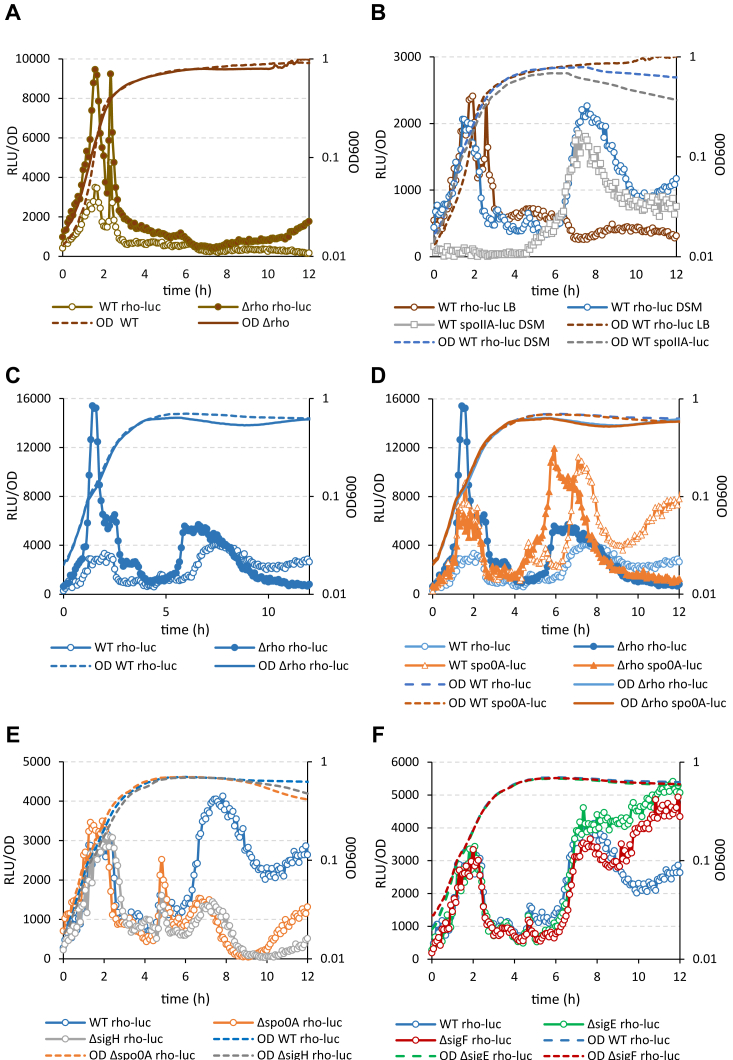
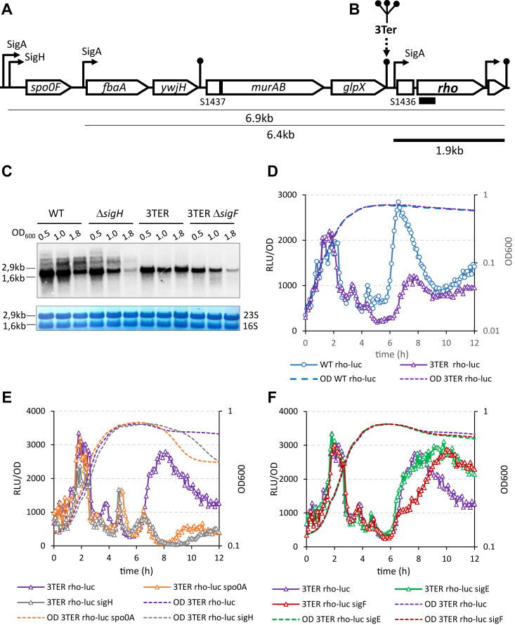
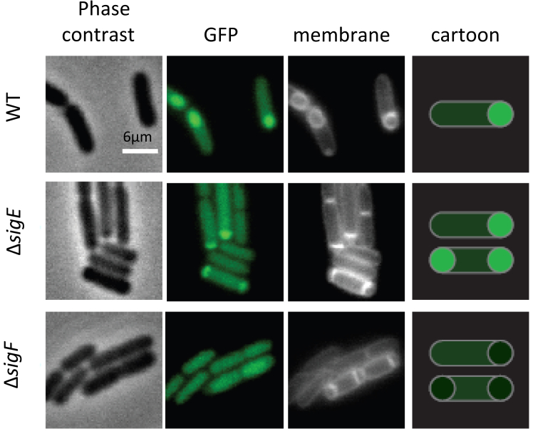
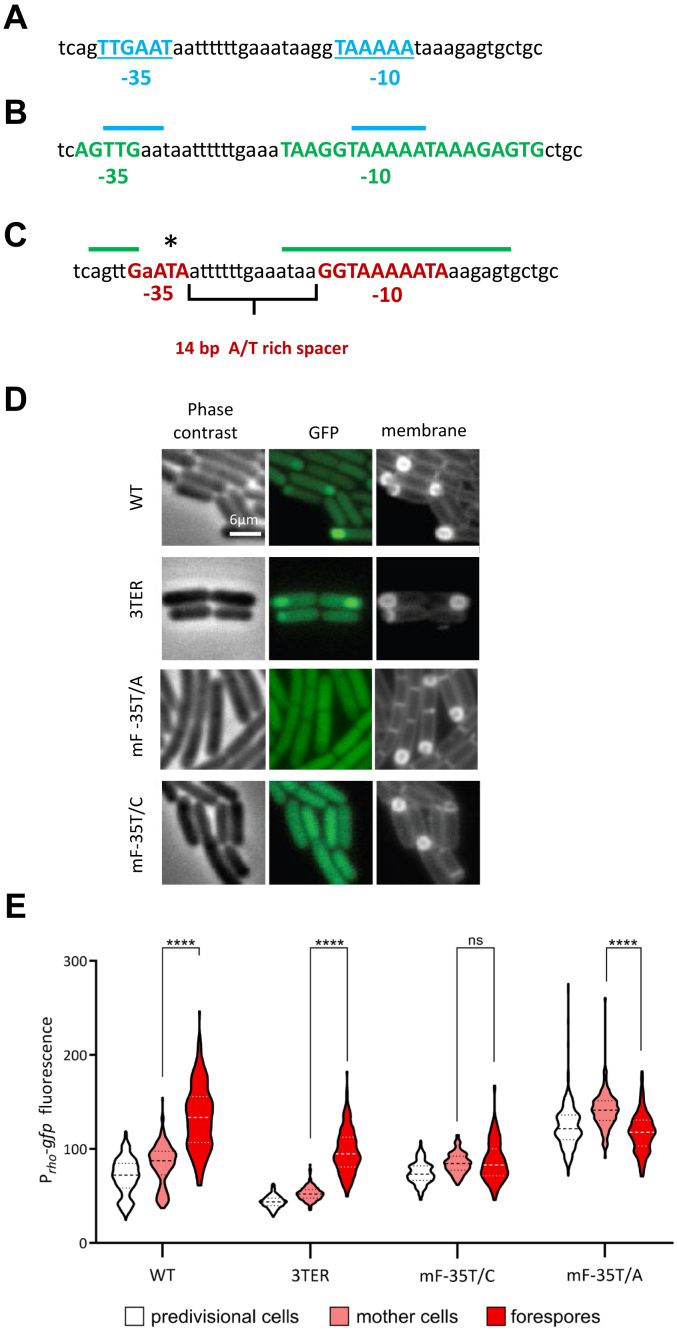
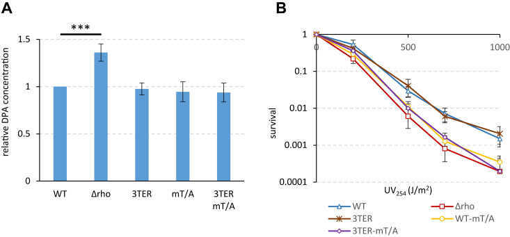
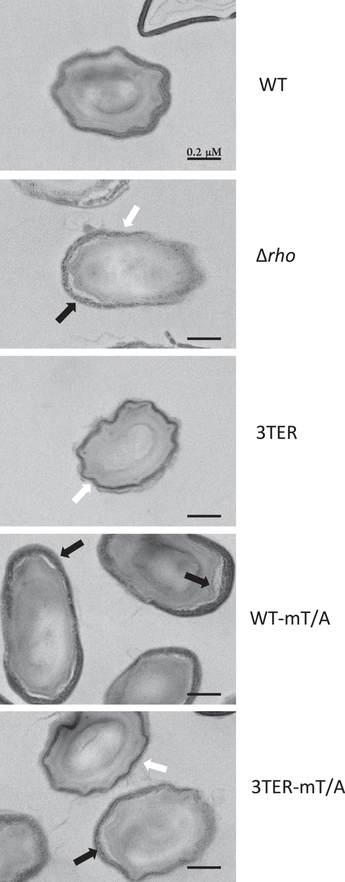
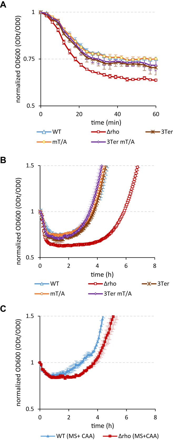
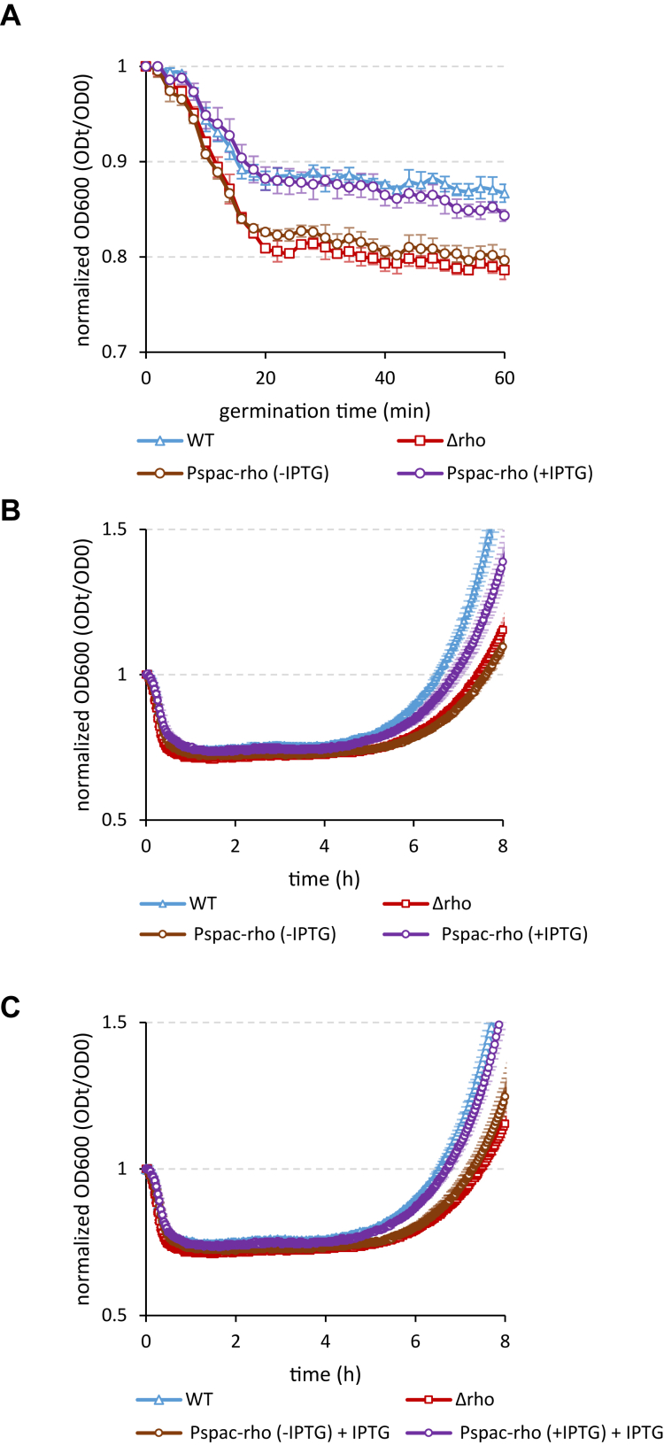
Termination factor Rho, responsible for the main factor-dependent pathway of transcription termination and the major inhibitor of antisense transcription, is an emerging regulator of various physiological processes in microorganisms. In Gram-positive bacterium Bacillus subtilis, Rho is involved in the control of cell adaptation to starvation and, in particular, in the control of sporulation, a complex differentiation program leading to the formation of a highly resistant dormant spore. While the initiation of sporulation requires a decrease in Rho protein levels during the transition to stationary phase, the mechanisms regulating the expression of rho gene throughout the cell cycle remain largely unknown. Here we show that a drop in the activity of the vegetative SigA-dependent rho promoter causes the inhibition of rho expression in stationary phase. However, after the initiation of sporulation, rho gene is specifically reactivated in two compartments of the sporulating cell using distinct mechanisms. In the mother cell, rho expression occurs by read-through transcription initiated at the SigH-dependent promoter of the distal spo0F gene. In the forespore, rho gene is transcribed from the intrinsic promoter recognized by the alternative sigma factor SigF. These regulatory elements ensure the activity of Rho during sporulation, which appears important for the proper formation of spores. We provide experimental evidence that disruption of the spatiotemporal expression of rho during sporulation affects the resistance properties of spores, their morphology, and the ability to return to vegetative growth under favorable growth conditions.

Keywords: Bacillus; Gram-positive bacteria; cell differentiation; gene expression; sporulation; transcription factor; transcription regulation; transcription termination.


Conflict of interest statement

Conflict of interest The authors declare that they have no conflicts of interest with the contents of this article.

Figures

Figure 1
Figure 1
Rho is specifically expressed during Bacillus subtilis sporulation.A, Rho expression in vegetative cells is limited to the exponential growth phase and autoregulated. Kinetics of luciferase activity in B. subtilis WT Prho-luc (empty circles) and Δrho Prho-luc (filled-in circles) cells grown in rich medium LB. In this and other panels, the plain dotted and solid lines represent growth curves of the Rho-proficient strains and rho-deletion mutant (Δrho), respectively, measured by A600. B, in the sporulation-inducing conditions, rho expression is additionally activated in stationary phase. Comparative analysis of the luciferase activity in WT Prho-luc cells grown in LB (brown circles) and in sporulation medium DSM (blue circles). Activation of luciferase expression from the early sporulation promoter spoIIA in the control WT PspoIIA-luc cells grown in DSM (gray squares) marks the initiation of sporulation. C, in the sporulation-inducing conditions, the autoregulation of Rho is weakened in stationary phase. Kinetics of luciferase activity in B. subtilis WT Prho-luc (empty blue circles) and Δrho Prho-luc (filled-in blue circles) cells grown in sporulation medium DSM. D, Rho expression in the sporulation-inducing conditions correlates with the activation of Spo0A. Comparative analysis of luciferase expression from Prho-luc (blue circles) and Pspo0A-luc (orange triangles) transcriptional fusions in WT (empty symbols) and Δrho (filled-in symbols) cells grown in DSM. E and F, Rho expression in stationary phase depends on the initiation of sporulation. Kinetics of luciferase expression from Prho-luc transcriptional fusions in WT cells (blue circles) and the sporulation mutants (E): Δspo0A (orange circles) and ΔsigH (gray circles); and (F): ΔsigF (red circles) and ΔsigE (green circles). Measurements were taken every 5 min after cells inoculation in media at optical density A600 ∼0.025 (time point 0). For each strain, plotted are the mean values of luminescence readings corrected for OD from four independent cultures analyzed simultaneously. Each panel presents the simultaneously collected data. The data in (C) and (D) and in (E) and (F) were obtained in two independent experiments. Each strain and growth condition was tested at least three times. The results from the representative experiment are presented.
Figure 2
Figure 2
Role of the read-through transcription in the rho expression during sporulation.A, schematic representation of gene organization and transcription within the rho locus of B. subtilis chromosome. Arrow-shaped and flat rectangles indicate protein-coding genes and noncoding RNA S-segments (11), respectively. Curved arrow-ended lines represent promoters; their regulatory sigma factors are indicated. Straight dot-ended lines represent intrinsic terminators. The underlying lines indicate the transcripts of the rho gene, which were identified in (11); the transcript initiated at the known rho promoter is bolded. The approximate size of the transcripts is shown. Small black rectangle schematizes the 5′ rho-specific riboprobe used in the Northern blot. B, cartoon of the insertion of three intrinsic terminators at the end of glpX gene in the 3TER strain. C, Northern blot analysis of the rho-specific transcripts in the WT and the mutant ΔsigH, 3TER, and 3TER ΔsigF strains during sporulation. Cells were grown in DSM and sampled during exponential growth (A600 0.5), the transition to stationary phase (A600 1.0), and in stationary phase (A600 1.8). Total RNA was extracted, processed, and hybridized with the rho-specific riboprobe as described in Experimental procedures. Upper panel. The rho-specific transcripts visualized by Northern blotting. The lines relate the position of the ribosomal 16S and 23S RNAs visualized by staining of the membrane with 0.2% methylene blue (bottom panel). The approximate size of the ribosomal RNAs is indicated. Bottom panel. Staining of the ribosomal 16S and 23S RNAs with methylene blue allows controlling the equilibrium of the loaded RNA samples and their transfer onto membrane. D, suppression of the read-through transcription inhibits rho expression in stationary phase. Kinetics of luciferase activity in B. subtilis WT Prho-luc (blue circles) and 3TER Prho-luc (violet triangles) cells grown in the sporulation-inducing DSM. E and F, in the absence of read-through transcription, the residual expression of rho in stationary phase is sporulation-dependent. Kinetics of luciferase activity in 3TER Prho-luc strain (violet triangles) and its sporulation mutants (E): Δspo0A (orange triangles) and ΔsigH (gray triangles); and (F): ΔsigF (red triangles) and ΔsigE (green triangles) grown in DSM. The data in (E) and (F) were obtained in the same experiment and are independent from (D). The data were collected and processed as described in Fig. 1. The experiments were reproduced at least three times. The results from the representative experiment are presented.
Figure 3
Figure 3
The expression of rho in the forespore compartment of sporangium depends on Sigma F. The WT Prho-gfp strain and its ΔsigF and ΔsigE mutant derivatives were induced for sporulation by the resuspension method as described in Experimental procedures. Cells were sampled 3 hours after resuspension in the nutrient-poor SM medium and observed by phase contrast microscopy and by epifluorescence illumination of GFP or membrane-affine dye in two independent replicas. The right-hand cartoon depicts spatial GFP-mediated fluorescence in cells with asymmetric septa.
Figure 4
Figure 4
Forespore-specific expression of rho depends on the activity of a genuine SigF-dependent promoter.A and B, sequence and structural elements of the SigA-dependent rho promoter reported by: (47) in (A) and (11) in (B). The −35 and −10 boxes are bolded and colored in blue (A) and green (B). In (B), the upper blue lines indicate the relative position of the rho promoter identified by (47). C, sequence and structural elements (bolded and colored in red) of putative SigF-dependent rho promoter identified in this analysis as matching consensus sequences recognized by SigF-containing RNA polymerase (44). The upper green lines indicate the relative position of the SigA-dependent rho promoter identified by (11). The asterisk indicates a conserved thymine nucleotide in the −35 sequence of the SigF-dependent promoter subjected to mutagenesis. D and E, mutations of the SigF-dependent rho promoter specifically inhibit rho expression in the forespores. The WT and 3TER cells bearing the nonmodified Prho-gfp fusion and WT Prho-gfp cells containing the indicated single-nucleotide mutations of putative SigF-dependent rho promoter were induced for sporulation and analyzed for GFP-mediated fluorescence as described in Experimental procedures and Fig. 3. D, Micrographs of typical cells observed by phase contrast, epifluorescence illumination of GFP, or membrane-affine dye. E, average fluorescence intensities, determined in predivisional cells (white) and the mother cell (rose) and the forespore (red) compartments of sporangia in each strain, are displayed as violin plots. Means and quartiles are represented as dashed and dotted lines, respectively. N > 100, per strain and per replica. Displayed is a representative experiment from two independent replica. The statistical significance was estimated by a two-tailed t test. p-values are displayed as follows: ∗∗∗∗ = p < 0.0001; ns = p > 0.
Figure 5
Figure 5
The alteration of the spatiotemporal expression of rho affects the resistance properties of mature spores. Spores produced by WT and 3TER cells expressing rho from the nonmodified promoters, their mutant derivatives WT-mT/A and 3TER-mT/A inactivated for rho expression in the forespore, and Δrho cells expressing no Rho were analyzed for the levels of dipicolinic acid (A) and the resistance to ultraviolet irradiation (B) as described in Experimental procedures. A, spore DPA contents are normalized to the WT level. The assay was reproduced 10 times with two independently prepared sets of five spores. Plotted are mean values from all measurements. Statistical significance was estimated with a two-tailed t test. The displayed p-value is as follows: ∗∗∗p ≤ 0.001. B, UV test was reproduced five times with one set of spores. The bars represent SD from the mean values.
Figure 6
Figure 6
The alteration of the spatiotemporal expression of rho affects the morphology of spores. Thin section transmission electron micrographs of spores produced by cells differentially expressing rho. White arrows indicate a thinner and less electron-dense outer coat in Δrho, 3TER, and 3TER-mT/A spores, which did not express rho in the forespores during maturation. Black arrows indicate the detachment of outer and inner coats in Δrho, WT-mT/A, and 3TER-mT/A spores, which did not express rho in the mother cells.
Figure 7
Figure 7
Rho activity determines the revival properties of spores. Spores of the WT (blue triangles), Δrho (red squares), 3TER (brown crosses), WT-mT/A (orange circles), and 3TER-mT/A (violet diamonds) strains were induced by the germinant L-alanine (10mM) and compared for germination (A) and outgrowth in the nutrient-poor MS medium (B) as described in Experimental procedures. C, WT (filled-in blue triangles) and Δrho (filled-in red squares) spores were germinated as in (A) and analyzed for outgrowth in the nutrient-replenished MS medium containing 0.5% casamino acids. The experiments were performed at least twice with three independent sets of spores. Each experiment included up to six replicas of individual suspensions of spores. The results of the representative experiment are plotted. The bars represent SD from the mean values.
Figure 8
Figure 8
The WT rate of spore outgrowth requires the expression of rho during spore morphogenesis and de novo after germination. Spores of the WT Pspac-rho strain expressing Rho protein from the IPTG-inducible promoter Pspac were produced in the absence (brown circles) or presence (violet circles) of IPTG 100μM and compared with the WT (blue triangles) and Δrho (red squares) spores for germination with L-alanine (A) and outgrowth in MS medium without (B) or with IPTG 100mM (C). Of note, induction of rho expression by IPTG (C) slightly accelerates the outgrowth compared to (B). The experiments were reproduced at least twice with two independent samples of the IPTG-induced or noninduced WT Pspac-rho spores. Each experiment included up to six replicas of individual suspensions of spores. The results of the representative experiment are plotted. The bars represent SD from the mean values.

References

    1. Peters J.M., Vangeloff A.D., Landick R. Bacterial transcription terminators: the RNA 3′-end chronicles. J. Mol. Biol. 2011;412:793–813. - PMC - PubMed
    1. Ray-Soni A., Bellecourt M.J., Landick R. Mechanisms of bacterial transcription termination: all good things must end. Annu. Rev. Biochem. 2016;85:319–347. - PubMed
    1. Kriner M.A., Sevostyanova A., Groisman E.A. Learning from the leaders: gene regulation by the transcription termination factor Rho. Trends Biochem. Sci. 2016;41:690–699. - PMC - PubMed
    1. Turnbough C.L., Jr. Regulation of bacterial gene expression by transcription attenuation. Microbiol. Mol. Biol. Rev. 2019;83:10–1128. - PMC - PubMed
    1. Mandell Z.F., Zemba D., Babitzke P. Factor-stimulated intrinsic termination: getting by with a little help from some friends. Transcription. 2022;13:96–108. - PMC - PubMed

Publication types

MeSH terms

LinkOut - more resources