. 2024 Oct 21;22(1):646.
doi: 10.1186/s12951-024-02877-8.

Alleviating rheumatoid arthritis with a photo-pharmacotherapeutic glycan-integrated nanogel complex for advanced percutaneous delivery

Affiliations

Alleviating rheumatoid arthritis with a photo-pharmacotherapeutic glycan-integrated nanogel complex for advanced percutaneous delivery

Pei-Wei Weng et al. J Nanobiotechnology. .

Abstract

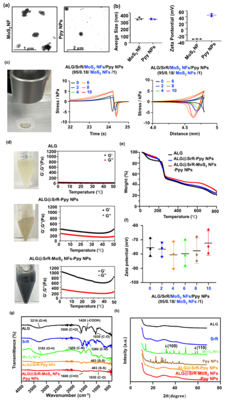
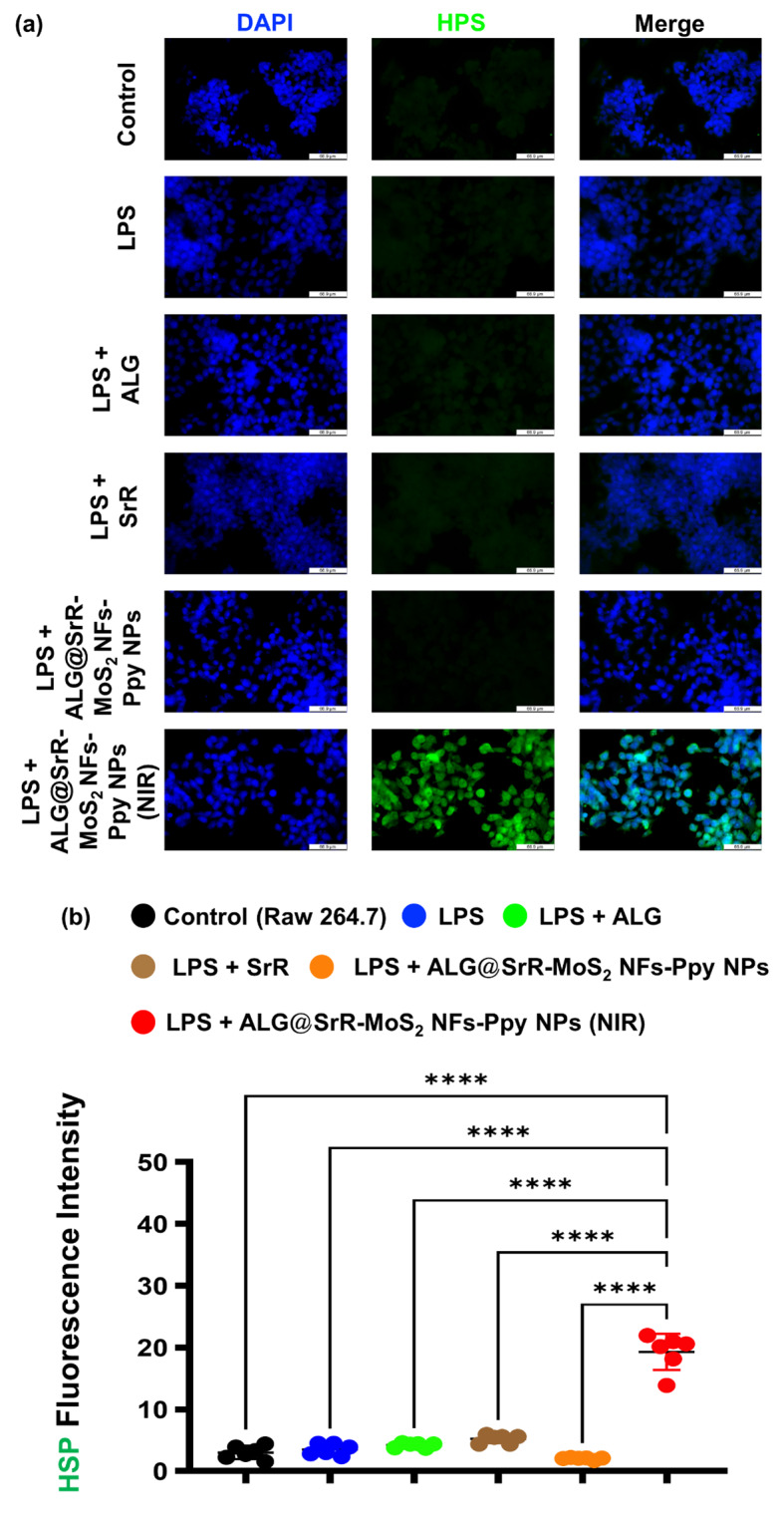
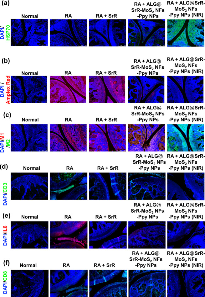
The prospective of percutaneous drug delivery (PDD) mechanisms to address the limitations of oral and injectable treatment for rheumatoid arthritis (RA) is increasing. These limitations encompass inadequate compliance among patients and acute gastrointestinal side effects. However, the skin's intrinsic layer can frequently hinder the percutaneous dispersion of RA medications, thus mitigating the efficiency of drug delivery. To circumvent this constraint, we developed a strontium ranelate (SrR)-loaded alginate (ALG) phototherapeutic hydrogel to assess its effectiveness in combating RA. Our studies revealed that this SrR-loaded ALG hydrogel incorporating photoelectrically responsive molybdenum disulfide nanoflowers (MoS2 NFs) and photothermally responsive polypyrrole nanoparticles (Ppy NPs) to form ALG@SrR-MoS2 NFs-Ppy NPs demonstrated substantial mechanical strength, potentially enabling delivery of hydrophilic therapeutic agents into the skin and significantly impeding the progression of RA. Comprehensive biochemical, histological, behavioral, and radiographic analyses in an animal model of zymosan-induced RA demonstrated that the application of these phototherapeutic ALG@SrR-MoS2 NFs-Ppy NPs effectively reduced inflammation, increased the presence of heat shock proteins, regulatory cluster of differentiation M2 macrophages, and alleviated joint degeneration associated with RA. As demonstrated by our findings, treating RA and possibly other autoimmune disorders with this phototherapeutic hydrogel system offers a distinctive, highly compliant, and therapeutically efficient method.

Keywords: Immunomodulation; Molybdenum disulfide; Phototherapy; Polypyrrole; Rheumatoid arthritis.


Conflict of interest statement

The authors affirmed that they have no competing interests to declare.

Figures

Scheme 1
Scheme 1
Schematic diagram demonstrating the innovative approach to synthesize composite biomaterials by integrating polypyrrole nanoparticles (Ppy NPs) and molybdenum disulfide nanoflowers (MoS2 NFs) into an alginate (ALG) hydrogel system to construct ALG@SrR-MoS2 NFs-Ppy NPs. The biomimetic combination of Ppy NPs and MoS2 NFs improved the mechanical strength of the gel by interacting in a coordinated manner and showed impressive photothermal and photoelectric conversion characteristics
Fig. 1
Fig. 1
A comprehensive overview of the fabrication and characterization of strontium ranelate incorporated into alginate-based hydrogels containing molybdenum disulfide nanoflowers and polypyrrole nanoparticles (ALG@SrR-MoS2 NFs-Ppy NPs). (a) Transmission electron microscopic (TEM) images provided detailed visualization of the morphologies of MoS2 NFs and Ppy NPs. (b) Dynamic light scattering (DLS) tests were performed to evaluate the hydrodynamic sizes of MoS2 NFs and Ppy NPs. (c) A texture analysis (TA) was conducted to examine the mechanical properties of formulations comprising ALG, SrR, MoS2 NF, and Ppy NPs in specific ratios (95/0.18/0/1, 95/0.18/2/1, 95/0.18/4/1, 95/0.18/6/1, 95/0.18/8/1, and 95/0.18/10/1). (d) Rheological experiments were carried out to assess the viscoelastic properties of ALG, ALG@SrR-Ppy NPs, and ALG@SrR-MoS2 NFs-Ppy NPs. (e) Thermogravimetric analysis (TGA) tests determined the thermal stability and decomposition characteristics of ALG@SrR-MoS2 NFs-Ppy NPs. (f) Zeta potential measurements were performed to understand the surface charge properties of the formulations with varying ratios of ALG, SrR, MoS2 NFs, Ppy NPs. (g) A Fourier transform infrared (FTIR) spectroscopic analysis provided insights into the chemical structures of and bonding among ALG, SrR, MoS2 NFs, ALG@SrR-Ppy NPs, and ALG@SrR-MoS2 NFs-Ppy NPs. (h) X-Ray diffraction (XRD) assessments offered information on the crystalline or amorphous nature of ALG, SrR, MoS2 NFs, Ppy NPs, ALG@SrR-Ppy NPs, and ALG@SrR-MoS2 NFs-Ppy NPs. These analyses collectively contributed to a deeper understanding of the physical, chemical, and thermal properties of the ALG@SrR-MoS2 NF-Ppy NP hydrogel, paving the way for potential biomedical applications
Fig. 2
Fig. 2
Findings of the photoresponsivity, encompassing both photothermal and photoelectric responses of various materials. (a) Photothermal results illustrate the thermal behavior of alginate (ALG), strontium ranelate (SrR), molybdenum disulfide nanoflowers (MoS2 NFs), polypyrrole nanoparticles (Ppy NPs), ALG@strontium ranelate (SrR)-Ppy NPs and the ALG@strontium ranelate (SrR)-MoS2 NF-Ppy NP composite. These were assessed under repeated near-infrared (NIR) irradiation (808 nm, 1.0 W/cm²) to evaluate their heat-generation capabilities. (b) Photoelectric results demonstrated the electrical responses of the same materials under similar conditions of frequent NIR irradiation. This part of the study focused on examining changes in electrical conductivity and the photoelectric efficiency during on-off cycles of NIR exposure. Together, these results offer comprehensive insights into the photothermal and photoelectric properties of ALG@SrR-MoS2 NFs-Ppy NPs, showcasing their potential applications in fields where photoresponsive behavior is crucial. Some study’s quantitative data are presented as the average ± standard deviation (SD) of a minimum of three replicate experiments (n ≥ 3)
Fig. 3
Fig. 3
SEM-EDS analysis of (a) alginate-incorporated strontium ranelate-polypyrrole nanoparticle (ALG@SrR-Ppy NPs) and (b) ALG@SrR-molybdenum disulfide nanoflower (MoS2 NFs)-Ppy NPs hydrogels. High-resolution SEM images illustrating microstructural differences between ALG@SrR-Ppy NPs and ALG@SrR-MoS2 NFs-Ppy NPs, with the latter displaying a regular and distinct porous morphology. EDS spectra highlighting the elemental composition of the hydrogels, with ALG@SrR-Ppy NPs showing a predominance of oxygen (O), carbon (C), nitrogen (N), and strontium (Sr). ALG@SrR-MoS2 NFs-Ppy NPs revealed similar elemental contents with the addition of sulfur (S) and molybdenum (Mo), indicating the successful incorporation of MoS2 NFs into the hydrogel nerk. The figure demonstrates the homogeneity and distinct structures within the hydrogel composites, validating their potential application in targeted drug delivery and regenerative medicine
Fig. 4
Fig. 4
In vitro investigation of the cellular viability and apoptosis in RAW 264.7 cells. (a) MTT assay results revealed the biocompatibility of the samples, indicating minimal cytotoxicity across all tested formulations including alginate (ALG), polypyrrole (Ppy) nanoparticles (NPs), molybdenum disulfide (MoS2) nanoflowers (NFs), strontium ranelate (SrR), and ALG@SrR-MoS2 NFs-Ppy NPs, both with and without near-infrared (NIR) irradiation. ALG/SrR/MoS2 NFs/Ppy NPs had a weight ratio of 95/0.18/6/1. (b) Fluorescence microscopic images demonstrating apoptosis using a calcein-AM/ethidium homodimer assay. Live cells are indicated by green fluorescence, while apoptotic cells are marked by red fluorescence. The group treated with ALG@SrR-MoS2 NFs-Ppy NPs exhibited a slight increase in red fluorescence, suggesting marginal apoptosis with the majority of cells remaining viable. Statistical significance thresholds were established at p values of < 0.05 (*), < 0.01 (**), and < 0.001 (***)
Fig. 5
Fig. 5
In vitro investigation of the cellular reactive oxygen species (ROS) levels in RAW 264.7 cells. (a, b) Data depicting levels of intracellular ROS after 24 h of incubation with different formulations, with a subset of cells stimulated by lipopolysaccharide (LPS). An Amplex red fluorescent probe was used for ROS quantification. Cells treated with LPS exhibited higher ROS levels, while those treated with the hydrogel formulations show reduced ROS, particularly in the ALG@SrR-MoS2 NF-Ppy NP + NIR group, which demonstrated a significant reduction in ROS (p < 0.01) after LPS stimulation, underscoring the potential anti-inflammatory effects of the phototherapeutic components. These data confirmed the protective effects of the ALG@SrR-MoS2 NF-Ppy NP hydrogel against cellular stress and highlighted the synergistic benefits of NIR irradiation in reducing inflammatory markers in macrophages. The study’s quantitative data are presented as the average ± standard deviation (SD) of a minimum of three replicate experiments (n ≥ 3). We used the two-way analysis of variance (ANOVA) to determine the statistical significance among several groups. GraphPad Prism software vers. 5.04 for Windows was used to do this analysis (Dotmatics, Boston, MA, USA). Thresholds of statistical significance were set to p < 0.05 (*), p < 0.01 (**), p < 0.001 (***), and p < 0.0001 (****)
Fig. 6
Fig. 6
In vitro investigation of the cellular immunomodulation in RAW 264.7 cells. (a) Fluorescent labeling microscopic data and (b) analysis of macrophage polarization. LPS-treated RAW 264.7 cells exhibited increased M1 phenotype markers (CD86, red fluorescence) and decreased M2 markers (CD206, green fluorescence). Treatment with ALG@SrR-MoS2 NFs-Ppy NPs + NIR resulted in a notable decrease in M1 marker expression and an increase in M2 marker expression, compared to groups of RAW 264.7 cells + LPS, RAW 264.7 cells + LPS + ALG, RAW 264.7 cells + LPS + SrR, and RAW 264.7 cells + LPS + ALG@SrR-MoS2 NFs-Ppy NPs, indicating a shift towards anti-inflammatory M2 polarization. The study’s quantitative data are presented as the average ± standard deviation (SD) of a minimum of three replicate experiments (n ≥ 3). We used the two-way analysis of variance (ANOVA) to determine the statistical significance among several groups. GraphPad Prism software vers. 5.04 for Windows was used to do this analysis (Dotmatics, Boston, MA, USA). Thresholds of statistical significance were set to p < 0.05 (*), p < 0.01 (**), p < 0.001 (***), and p < 0.0001 (****)
Fig. 7
Fig. 7
In vitro investigation of the cellular heat shock protein (HSP) levels in RAW 264.7 cells. (a) Microscopic and (b) quantitative evaluation of HSP expression by RAW 264.7 cells. Cells treated with LPS and ALG@SrR-MoS2 NFs-Ppy NPs + NIR demonstrated enhanced HSP expression compared to groups of RAW 264.7 cells + LPS, RAW 264.7 cells + LPS + ALG, RAW 264.7 cells + LPS + SrR, and RAW 264.7 cells + LPS + ALG@SrR-MoS2 NFs-Ppy NPs suggesting a protective effect against cellular stress. The study’s quantitative data are presented as the average ± standard deviation (SD) of a minimum of three replicate experiments (n ≥ 3). We used the two-way analysis of variance (ANOVA) to determine the statistical significance among several groups. GraphPad Prism software vers. 5.04 for Windows was used to do this analysis (Dotmatics, Boston, MA, USA). Thresholds of statistical significance were set to p < 0.05 (*), p < 0.01 (**), p < 0.001 (***), and p < 0.0001 (****)
Fig. 8
Fig. 8
Evaluation of drug penetration, immune response, and functional recovery in rheumatoid arthritis (RA) mice treated with phototherapeutic hydrogels. (a, b) Fluorescein isothiocyanate (FITC) fluorescence imaging and analysis demonstrate the biodistribution of strontium ranelate (SrR) + FITC and SrR incorporated into alginate-based hydrogels containing molybdenum disulfide nanoflowers and polypyrrole nanoparticles (ALG@SrR-MoS2 NFs-Ppy NPs) containing FITC, with and without near infrared (NIR) treatment. Images reveal that the ALG@SrR-MoS2 NF-Ppy NP + NIR group achieved significant fluorescence in the joint cavity, indicating enhanced drug delivery. (c, d) Microscopic and quantitative analyses of Cy5 skin penetration and macrophage targeting in RA mice treated with SrR + Cy5 + NIR versus ALG@SrR-MoS2 NFs-Ppy NPs containing Cy5 + NIR. The latter group exhibited increased Cy5 fluorescence and macrophage accumulation, underscoring the targeted delivery and anti-inflammatory potential of the hydrogel. (e) Thermal imaging revealed that the ALG@SrR-MoS2 NFs-Ppy NPs exhibited a pronounced photothermal effect when subjected to NIR irradiation. (f, g) A gait analysis was used to measure the severity of arthritis and functional recovery. The highest stride frequency values were observed in the healthy control group, while the lowest were in the untreated RA group. Mice treated with ALG@SrR-MoS2 NFs-Ppy NPs + NIR showed an improved stride frequency, indicating alleviation of arthritic symptoms and enhanced mobility. These findings collectively demonstrated the profound impact of ALG@SrR-MoS2 NF-Ppy NP + NIR treatment on drug penetration, immune modulation, and functional improvement in this RA mouse model. The study’s quantitative data are presented as the average ± standard deviation (SD) of a minimum of three replicate experiments (n ≥ 3). We used the two-way analysis of variance (ANOVA) to determine the statistical significance among several groups. GraphPad Prism software vers. 5.04 for Windows was used to do this analysis (Dotmatics, Boston, MA, USA). The Student’s t-test was used for the statistical analysis of Fig. 8d. Thresholds of statistical significance were set to p < 0.05 (*), p < 0.01 (**), p < 0.001 (***), and p < 0.0001 (****)
Fig. 9
Fig. 9
Toxicity assessment, cartilage erosion analysis, and MRI evaluation in rheumatoid arthritis (RA) mice treated with strontium ranelate incorporated into alginate-based hydrogels containing molybdenum disulfide nanoflowers and polypyrrole nanoparticles (ALG@SrR-MoS2 NFs-Ppy NPs). (a) H&E staining of major organs (heart, liver, spleen, lungs, and kidneys) from different treatment groups, including ALG@SrR-MoS2 NFs-Ppy NPs with near infrared (NIR) therapy. Staining results indicated no significant morphological changes in these organs compared to the sham group, suggesting minimal systemic toxicity. (b) Alcian blue (AB) staining of knee joint cartilage in various groups. Light-blue staining in untreated and SrR-treated arthritic groups indicates potential cartilage damage, while intense staining in the ALG@SrR-MoS2 NF-Ppy NP + NIR group resembles healthy cartilage, indicating effective protection against cartilage erosion. (c) T2-weighted MRI images were used to assess cartilage thickness and tissue integrity in RA knees. This comprehensive analysis underscores the safety and efficacy of ALG@SrR-MoS2 NFs-Ppy NPs, particularly when combined with NIR therapy, in mitigating arthritis symptoms and progression, while maintaining a favorable safety profile. Quantitative data in this study are expressed as the mean ± standard deviation of at least three experiments done in triplicate (n ≥ 3). To ascertain the statistical significance between multiple groups, we employed the nonparametric Kruskal-Wallis ANOVA. This analysis was conducted using GraphPad Prism software vers. 5.04 for Windows. Statistical significance thresholds were established at p values of < 0.05 (*), < 0.01 (**), < 0.001 (***), and 0.0001 (****)
Fig. 10
Fig. 10
Assessment of heat shock protein (HSP) levels, reactive oxygen species (ROS), interleukin (IL)-6, T-cell activity (CD8+), T-cell activity (CD3+), and macrophage polarization in rheumatoid arthritis (RA) mice treated with strontium ranelate incorporated into alginate-based hydrogel containing molybdenum disulfide nanoflowers and polypyrrole nanoparticles (ALG@SrR-MoS2 NFs-Ppy NPs). (a) HSP expression in cartilage tissues, showing elevated HSP levels in the ALG@SrR-MoS2 NFs-Ppy NPs + NIR group, suggesting enhanced protective effects on cartilage lesions and potential applications in tissue engineering. (b) ROS detection using an Amplex red assay. Elevated ROS levels in the arthritis control group indicated increased inflammation, while a significant reduction in ROS was observed in groups treated with ALG@SrR-MoS2 NFs-Ppy NPs, especially with additional NIR therapy, indicating effective anti-inflammatory action. (c) Immunofluorescence (IF) staining for the macrophage polarization analysis in synovial tissues. Elevated CD86 expression (an M1 macrophage marker) in the RA control and treatment groups without NIR indicates a predominance of proinflammatory M1 macrophages. In contrast, the ALG@SrR-MoS2 NF-Ppy NP + NIR group showed reduced CD86 expression and increased CD206 expression (an M2 macrophage marker), highlighting effective modulation towards anti-inflammatory M2 macrophages. (d) Cellular expressions of CD3+ and (e) IL-6 in the treated groups. (f) CD8+ in the treated groups. These findings comprehensively demonstrate the multifaceted therapeutic impacts of ALG@SrR-MoS2 NF-Ppy NP + NIR treatment in an RA model, encompassing enhanced HSP expression, reduced ROS levels, and favorable macrophage polarization, thereby substantiating its potential for clinical application in RA management.

References

    1. Smolen JS, Aletaha D, McInnes IB. Rheumatoid arthritis. Lancet. 2016;388:2023–38. 10.1016/S0140-6736(16)30173-8. - PubMed
    1. Combe B, Landewe R, Daien CI, Hua C, Aletaha D, Álvaro-Gracia JM, Bakkers M, Brodin N, Burmester GR, Codreanu C, et al. 2016 update of the EULAR recommendations for the management of early arthritis. Ann Rheum Dis. 2017;76:948–59. 10.1136/annrheumdis-2016-210602. - PubMed
    1. Aletaha D, Smolen JS. Diagnosis and management of rheumatoid arthritis: a review. JAMA. 2018;320:1360–72. 10.1001/jama.2018.13103. - PubMed
    1. Wang W, Zhou H, Liu L. Side effects of methotrexate therapy for rheumatoid arthritis: a systematic review. Eur J Med Chem. 2018;158:502–16. 10.1016/j.ejmech.2018.09.027. - PubMed
    1. Ren S, Liu H, Wang X, Bi J, Lu S, Zhu C, Li H, Kong W, Chen R, Chen Z. Acupoint nanocomposite hydrogel for simulation of acupuncture and targeted delivery of triptolide against rheumatoid arthritis. J Nanobiotechnol. 2021;19:409. 10.1186/s12951-021-01157-z. - PMC - PubMed