. 2024 Oct 20;15(1):9055.
doi: 10.1038/s41467-024-53420-0.

Conformational cycle of a protease-containing ABC transporter in lipid nanodiscs reveals the mechanism of cargo-protein coupling

Affiliations

Conformational cycle of a protease-containing ABC transporter in lipid nanodiscs reveals the mechanism of cargo-protein coupling

Ruojing Zhang et al. Nat Commun. .

Abstract

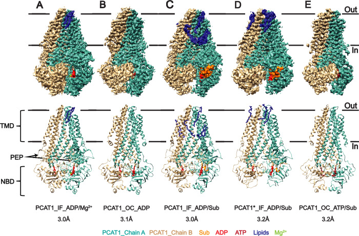
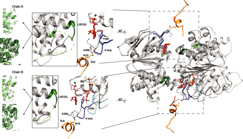
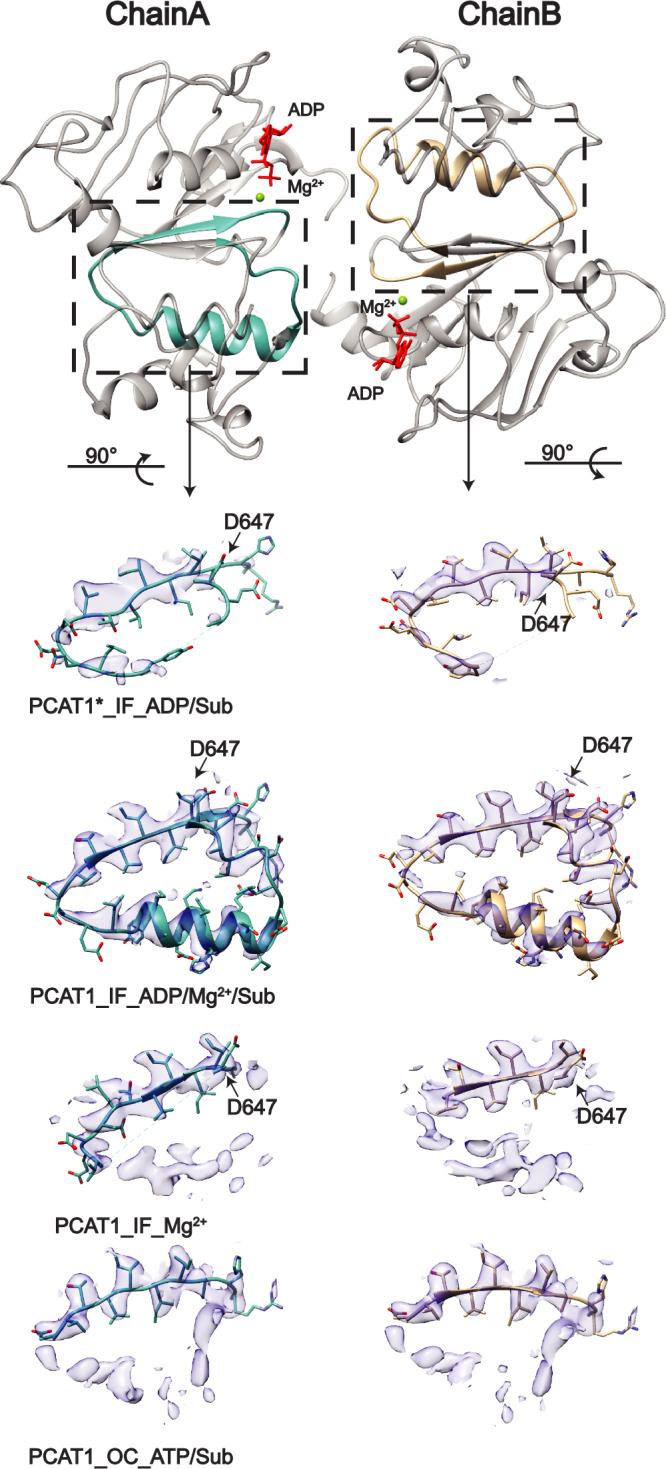
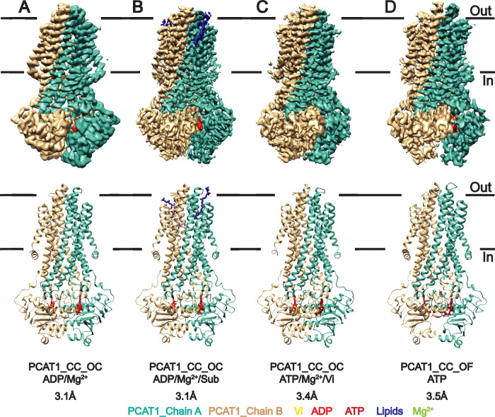
Protease-containing ABC transporters (PCATs) couple the energy of ATP hydrolysis to the processing and export of diverse cargo proteins across cell membranes to mediate antimicrobial resistance and quorum sensing. Here, we combine biochemical analysis, single particle cryoEM, and DEER spectroscopy in lipid bilayers along with computational analysis to illuminate the structural and energetic underpinnings of coupled cargo protein export. Our integrated investigation uncovers competitive interplay between nucleotides and cargo protein binding that ensures the latter's orderly processing and subsequent transport. The energetics of cryoEM structures in lipid bilayers are congruent with the inferred mechanism from ATP turnover analysis and reveal a snapshot of a high-energy outward-facing conformation that provides an exit pathway into the lipid bilayer and/or the extracellular side. DEER investigation of the core ABC transporter suggests evolutionary tuning of the energetic landscape to fulfill the function of substrate processing prior to export.


Conflict of interest statement

The authors declare no competing interests.

Figures

Fig. 1
Fig. 1. Interplay of ADP and Sub in the inhibition and stimulation of PCAT1 ATP turnover activity.
A Sub stimulates ATP hydrolysis whereas ADP competitively inhibits it (see Supplementary Table 1). Sub partly reverses the inhibition of ADP. PCAT1 turnover data is the average of 15 repeats from 8 biological repeats. PCAT1 inhibition with 2 mM ADP with 5 mM Vi is the average of 4 technical from 2 biological repeats. Turnover data with 5× Sub is derived from 5 biological repeats. B Biphasic shape of ATP turnover determined under 10 mM Mg2+ curve suggests partial inhibition by free Mg2+. The data is the average from at least 2 biological repeats. C Multiphasic dependence of Vmax on the Mg2+ to ATP ratio. The data is the average of at least two biological repeats. All data are presented as mean values ± standard deviation.
Fig. 2
Fig. 2. CryoEM structures of PCAT1 bound to nucleotides and Sub in lipid nanodiscs.
Both ADP (panel B) and ATP (panel E) stabilize an occluded (OC) conformation in the absence of Mg2+. Sub (panel C) and Mg2+ (panel A) shift the equilibrium toward the inward-facing (IF) conformation in the presence of ADP, whereas ATP/Sub bound PCAT1 remains in an OC conformation. PCAT1* denotes the cysteine-free PCAT1 (panel D).
Fig. 3
Fig. 3. Local structural changes induced by Sub binding.
Comparison of PCAT1_IF_ADP/Mg2+_class2 structure(light color) and PCAT1_ IF_ADP/Mg2+/Sub structure(dark color) shows that the leader peptide interacts with the A-loop altering its conformation. In addition, its binding stabilizes the signature motif as evidenced by the stronger density in that region in the IF structure determined in the presence of Sub (dark green). The cryoEM densities are shown at 4.6 σ for PCAT1_ IF_ADP/Mg2+/Sub and 7.7 σ for PCAT1_IF_ADP/Mg2+_class2.
Fig. 4
Fig. 4. Binding of nucleotides and Mg2+ stabilizes the 650–668 helix.
CryoEM densities are shown at the 0.09 contour level in Chimera. The cryo-EM maps are shown at 2.4 σ, 1.8 σ, 1.5 σ, 1.7 σ from top to bottom.
Fig. 5
Fig. 5. CryoEM structures of the ABC transporter core of PCAT1, PCAT1_CC.
Unlike the full length PCAT1, binding of Mg2+ (panel A, panel B with sub bound, panel C with ATP and Vi bound) does not stabilize the IF conformation in the cryoEM grids. ATP in the absence of Mg2+ stabilizes the OF conformation (panel D).
Fig. 6
Fig. 6. Structure of putative OF conformation of PCAT1 in lipid Nanodsics.
Comparison of the putative OF conformation of PCAT1 in detergent micelles (gold) with the OF conformation of the transporter core, PCAT1_CC, in lipid nanodiscs (teal). A Side view of the two structures highlighting the opening of the latter to the outer leaflet of the membrane. B Extracellular view identifying the movement of individual helices.
Fig. 7
Fig. 7. DEER analysis of the conformational dynamics of the core transporter in lipid nanodiscs.
Experimental and predicted distance distributions are shown in solid and dashed lines respectively. Binding of ATP-γS (solid green traces) induces large distance changes in the NBDs and the intracellular side of the TMD. OF_8VP1 is the OF from this paper.
Fig. 8
Fig. 8. DEER analysis of the conformational dynamics of the core transporter.
DEER distance distributions of the core transporter in lipid nanodiscs under turnover conditions (solid teal traces). Experimental and predicted distance distributions are shown in solid and dashed lines respectively. Except for 307 and 498, the distributions under turnover conditions are superimposable with those bound to ADP/Mg2+ (solid purple traces).
Fig. 9
Fig. 9. Model of PCAT1 conformational cycle.
The coupled cycle is initiated by binding of Sub either to the PCAT1 bound to ATP/Mg2+ in the IF conformation (1a) or to ADP-bound PCAT1 in the OC conformation (1b). The coupled cycle proceeds through processing of Sub (2) followed by ATP hydrolysis (3) to populate an OF conformation that releases leaderless Sub to the extracellular side or the outer leaflet of the membrane. Subsequently, Mg2+ dissociation (4b) could trap the transporter in an occluded conformation to reduce futile ATP hydrolysis. Alternatively, Mg2+-ADP dissociation with Mg2+-ATP binding (4a) could facilitate an IF conformation in an uncoupled cycle without Sub binding where ATP hydrolysis (5a) followed by dissociation of Mg2+ (5b) results in the OC conformation.

References

    1. Thomas, C. & Tampe, R. Structural and mechanistic principles of ABC transporters. Annu Rev. Biochem.89, 605–636 (2020). - PubMed
    1. Locher, K. P. Mechanistic diversity in ATP-binding cassette (ABC) transporters. Nat. Struct. Mol. Biol.23, 487–493 (2016). - PubMed
    1. Thomas, C. et al. Structural and functional diversity calls for a new classification of ABC transporters. FEBS Lett.594, 3767–3775 (2020). - PMC - PubMed
    1. Gebhard, S. A. B. C. transporters of antimicrobial peptides in Firmicutes bacteria - phylogeny, function and regulation. Mol. Microbiol.86, 1295–1317 (2012). - PubMed
    1. Gebhard, S. & Mascher, T. Antimicrobial peptide sensing and detoxification modules: unravelling the regulatory circuitry of Staphylococcus aureus. Mol. Microbiol.81, 581–587 (2011). - PubMed

Publication types

MeSH terms

LinkOut - more resources