. 2024 Oct;13(10):e70004.
doi: 10.1002/jev2.70004.

Bacterial extracellular vesicles as intranasal postbiotics: Detailed characterization and interaction with airway cells

Affiliations

Bacterial extracellular vesicles as intranasal postbiotics: Detailed characterization and interaction with airway cells

Agnieszka Razim et al. J Extracell Vesicles. 2024 Oct.

Abstract

Escherichia coli A0 34/86 (EcO83) is a probiotic strain used in newborns to prevent nosocomial infections and diarrhoea. This bacterium stimulates both pro- and anti-inflammatory cytokine production and its intranasal administration reduces allergic airway inflammation in mice. Despite its benefits, there are concerns about the use of live probiotic bacteria due to potential systemic infections and gene transfer. Extracellular vesicles (EVs) derived from EcO83 (EcO83-EVs) might offer a safer alternative to live bacteria. This study characterizes EcO83-EVs and investigates their interaction with host cells, highlighting their potential as postbiotic therapeutics. EcO83-EVs were isolated, purified, and characterised following the Minimal Information of Studies of Extracellular Vesicles (MISEV) guidelines. Ex vivo studies conducted in human nasal epithelial cells showed that EcO83-EVs increased the expression of proteins linked to oxidative stress and inflammation, indicating an effective interaction between EVs and the host cells. Further in vivo studies in mice demonstrated that EcO83-EVs interact with nasal-associated lymphoid tissue, are internalised by airway macrophages, and stimulate neutrophil recruitment in the lung. Mechanistically, EcO83-EVs activate the NF-κΒ signalling pathway, resulting in the nitric oxide production. EcO83-EVs demonstrate significant potential as a postbiotic alternative to live bacteria, offering a safer option for therapeutic applications. Further research is required to explore their clinical use, particularly in mucosal vaccination and targeted immunotherapy strategies.

Keywords: EVs; Ec083; NF‐κΒ signalling; bacterial extracellular vesicles; macrophage; nitric oxide; postbiotics; probiotic.


Conflict of interest statement

The authors report no conflict of interest.

Figures

FIGURE 1
FIGURE 1
Production and purification of EcO83‐EVs. EcO83 was grown in a BHI medium for 8 h, the bacteria culture were spun down, and the supernatant was filtered through a 0.22 µm pore membrane. The filtrate was then ultracentrifuged and purified on the OptiPrep gradient. Collected fractions were ultracentrifuged, characterized and pooled to obtain pure EcO83‐EVs. Created with BioRender.
FIGURE 2
FIGURE 2
Characterization of EcO83‐EVs. (a) Zetasizer analysis depicting the mean particle concentration of EcO83‐EVs (n = 3). (b) Zetasizer particle size measurement of EcO83‐EVs (n = 3). (c) Nanoparticle Tracking Analysis of EcO83‐EVs. (d) Cryo‐EM visualization and quantification of EcO83‐EVs (scale bar = 50 nm). (e) TEM image of EcO83‐EVs (scale bar = 200 nm). (f) Left: representative AFM image of EcO83‐EVs (scale bar = 500 nm); green arrows—EVs, blue arrows—fibrillary structures, yellow arrows—amorphous structures, red arrows—deflated EVs. Right: contact angle versus diameter plot of 1760 individual particles found in EcO83‐EVs samples.
FIGURE 3
FIGURE 3
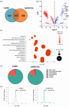
Proteomic and lipidomic analysis of EcO83‐EVs. (a) Venn diagram of proteins identified in EcO83‐EVs and EcO83 bacterial lysate. (b) Volcano plot of proteins upregulated (blue) and downregulated (red) when EcO83‐EVs were compared to bacterial lysate. (c) Bubble plot showing results of Fishers’ test comparing proteins upregulated in EcO83‐EVs against all identified proteins. (d) Lipidomic analysis of the EcO83 and EcO83‐EVs. (e) Zeta potential measurements of EcO83 and EcO83‐EVs.
FIGURE 4
FIGURE 4
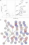
Proteomics of human nasal epithelial cells treated EcO83‐EVs ex vivo. (a) Volcano plot differentially expressed proteins from adult air‐liquid interface nasal epithelial cell cultures after 24 h, at 32°C incubation with 100 ng/mL EcO83‐EVs (according to protein content) compared to PBS‐treated cells. (b) STRING analysis of these differentially expressed proteins. Proteins that are upregulated are circled in blue, and downregulated proteins are shown in red.
FIGURE 5
FIGURE 5
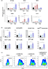
EcO83‐EVs interaction with mouse airways. Mice were administered rhodamine‐labelled or unlabelled 2.5 × 1010 EcO83‐EVs intranasally and sacrificed at either 0.5 h (= 5) or 2 h (n = 5) post‐administration. Control mice (= 3) were treated with 0.9% NaCl. The lungs and NALT were subsequently isolated for analysis. (a) Expression of specific genes at 0.5 and 2 h after treatment with unlabelled EcO83‐EVs analysed by qPCR. Reference gene α‐actin (ACTB) was used to normalize the gene expression data. (b) Cells recruited into lungs after 0.5 h or 2 h treatment with unlabelled EcO83‐EVs analysed by FACS. (c) FACS data obtained from lungs cells isolated from mice intranasally treated with rhodamine‐labelled EcO83‐EVs. Data shown for one representative experiment out of two. Statistical differences between samples were assessed using one‐way ANOVA; significance levels are denoted as *p ≤ 0.05, **p ≤ 0.01 ***p ≤ 0.001 ****p ≤ 0.0001 versus control.
FIGURE 6
FIGURE 6
EcO83‐EVs interaction with BMDMs. (a) Viability of WT BMDMs when treated with 1 × 109 and 1 × 1010 EcO83‐EVs/mL or 1 × 106 and 1 × 107 EcO83/mL for 24 h. (b) Production of NO in WT BMDMs when treated with 1 × 109 and 1 × 1010 EcO83‐EVs/mL or 1 × 106 and 1 × 107 EcO83/mL for 24 h. (c) NO production in TLR4 KO BMDMs treated with 1 × 109 and 1 × 1010 EcO83‐EVs/mL or 1 × 106 and 1 × 107 EcO83/mL for 24 h. (d) Production of NO in WT BMDMs when pre‐treated for 1 h with iNOS inhibitor (S‐MIU, 10 µM) and next treated with 1 × 109 and 1 × 1010 EcO83‐EVs/mL or 1 × 106 and 1 × 107 EcO83/mL for 24 h. (e) iNOS expression in WT BMDMs treated with 1 × 109 and 1 × 1010 EcO83‐EVs/mL or 1 × 106 and 1 × 107 EcO83/mL for 24 h, analysed by immunoblotting of cell lysates with monoclonal anti‐iNOS antibodies. Non‐treated cells (sham) were used as a negative control and LPS from E. coli O55B5 (1 µg/mL) as positive control for NO and iNOS induction. One‐way ANOVA was used to examine mean differences between samples. *p ≤ 0.05, **p ≤ 0.01 ***p ≤ 0.001 ****p ≤ 0.0001 versus control.
FIGURE 7
FIGURE 7
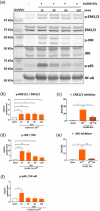
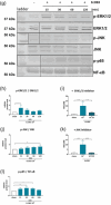
EcO83‐EVs and EcO83 effect on the MAPK and NF‐κΒ transcription factor activation in BMDM cells. (a) Phosphorylation status of ERK1/2, JNK, and p65 proteins in BMDMs treated with 1 × 1010 EcO83‐EVs/mL for 15–120 min analysed with immunoblotting. (b) Densitometry analysis of p‐ERK1/2/ERK1/2 in BMDM cells treated with 1 × 1010 EcO83‐EVs/mL for 15–120 min. (c) NO production of BMDM cells treated with 1 × 1010 EcO83‐EVs/mL for 24 h with/without 1 h pre‐treatment of ERK1/2 inhibitor (20 µM of U0126). (d) Densitometry analysis of p‐JNK/JNK in BMDM cells treated with 1 × 1010 EcO83‐EVs/mL for 15–120 min. (e) NO production of BMDM cells treated with 1 × 1010 EcO83‐EVs/mL for 24 h with/without 1 h pre‐treatment of JNK inhibitor (10 µM of SP600125). (f) Densitometry analysis of p‐p65/NF‐κΒ in BMDM cells treated with 1 × 1010 EcO83‐EVs/mL for 15–120 min. (g) Phosphorylation status of ERK1/2, JNK and p65 proteins in BMDMs treated with 1 × 107 EcO83/mL for 15–120 min analysed with immunoblotting. (h) Densitometry analysis of p‐ERK1/2/ERK1/2 in BMDM cells treated with 1 × 107 EcO83/mL for 15–120 min. (i) NO production of BMDM cells treated with 1 × 107 EcO83/mL for 24 h with/without 1 h pre‐treatment of ERK1/2 inhibitor (20 µM of U0126; the upstream kinase of p42 and p44 ERK1/2). (j) Densitometry analysis of p‐JNK/JNK in BMDM cells treated with 1 × 107 EcO83/mL for 15 ‐ 120 min. (k) NO production of BMDM cells treated with 1 × 107 EcO83/mL for 24 h with/without 1 h pre‐treatment of JNK inhibitor (10 µM of SP600125). (l) Densitometry analysis of p‐p65/NF‐κΒ in BMDM cells treated with 1 × 107 EcO83/mL for 15–120 min. One‐way ANOVA was used to examine mean differences between samples. *p ≤ 0.05, **p ≤ 0.01 ***p ≤ 0.001 ****p ≤ 0.0001 versus control.
FIGURE 7
FIGURE 7
EcO83‐EVs and EcO83 effect on the MAPK and NF‐κΒ transcription factor activation in BMDM cells. (a) Phosphorylation status of ERK1/2, JNK, and p65 proteins in BMDMs treated with 1 × 1010 EcO83‐EVs/mL for 15–120 min analysed with immunoblotting. (b) Densitometry analysis of p‐ERK1/2/ERK1/2 in BMDM cells treated with 1 × 1010 EcO83‐EVs/mL for 15–120 min. (c) NO production of BMDM cells treated with 1 × 1010 EcO83‐EVs/mL for 24 h with/without 1 h pre‐treatment of ERK1/2 inhibitor (20 µM of U0126). (d) Densitometry analysis of p‐JNK/JNK in BMDM cells treated with 1 × 1010 EcO83‐EVs/mL for 15–120 min. (e) NO production of BMDM cells treated with 1 × 1010 EcO83‐EVs/mL for 24 h with/without 1 h pre‐treatment of JNK inhibitor (10 µM of SP600125). (f) Densitometry analysis of p‐p65/NF‐κΒ in BMDM cells treated with 1 × 1010 EcO83‐EVs/mL for 15–120 min. (g) Phosphorylation status of ERK1/2, JNK and p65 proteins in BMDMs treated with 1 × 107 EcO83/mL for 15–120 min analysed with immunoblotting. (h) Densitometry analysis of p‐ERK1/2/ERK1/2 in BMDM cells treated with 1 × 107 EcO83/mL for 15–120 min. (i) NO production of BMDM cells treated with 1 × 107 EcO83/mL for 24 h with/without 1 h pre‐treatment of ERK1/2 inhibitor (20 µM of U0126; the upstream kinase of p42 and p44 ERK1/2). (j) Densitometry analysis of p‐JNK/JNK in BMDM cells treated with 1 × 107 EcO83/mL for 15 ‐ 120 min. (k) NO production of BMDM cells treated with 1 × 107 EcO83/mL for 24 h with/without 1 h pre‐treatment of JNK inhibitor (10 µM of SP600125). (l) Densitometry analysis of p‐p65/NF‐κΒ in BMDM cells treated with 1 × 107 EcO83/mL for 15–120 min. One‐way ANOVA was used to examine mean differences between samples. *p ≤ 0.05, **p ≤ 0.01 ***p ≤ 0.001 ****p ≤ 0.0001 versus control.

References

    1. Aderem, A. , & Ulevitch, R. J. (2000). Toll‐like receptors in the induction of the innate immune response. Nature, 406, 782–787. 10.1038/35021228 - DOI - PubMed
    1. Andrabi, S. M. , Sharma, N. S. , Karan, A. , Shahriar, S. M. S. , Cordon, B. , Ma, B. , & Xie, J. (2023). Nitric oxide: Physiological functions, delivery, and biomedical applications. Advanced Science, 10, 2303259. 10.1002/advs.202303259 - DOI - PMC - PubMed
    1. Atri, C. , Guerfali, F. Z. , & Laouini, D. (2018). Role of human macrophage polarization in inflammation during infectious diseases. International Journal of Molecular Sciences, 19, 1801. 10.3390/ijms19061801 - DOI - PMC - PubMed
    1. Bakkari, M. A. , Valiveti, C. K. , Kaushik, R. S. , & Tummala, H. (2021). Toll‐like receptor‐4 (TLR4) agonist‐based intranasal nanovaccine delivery system for inducing systemic and mucosal immunity. Molecular Pharmaceutics, 18, 2233–2241. 10.1021/acs.molpharmaceut.0c01256 - DOI - PubMed
    1. Bayarri, M. A. , Milara, J. , Estornut, C. , & Cortijo, J. (2021). Nitric oxide system and bronchial epithelium: More than a barrier. Frontiers in Physiology, 12, 687381. 10.3389/fphys.2021.687381 - DOI - PMC - PubMed

LinkOut - more resources