. 2024 Nov;55(11):2705-2715.
doi: 10.1161/STROKEAHA.124.048264. Epub 2024 Oct 21.

Perinatal Caffeine Administration Improves Outcomes in an Ovine Model of Neonatal Hypoxia-Ischemia

Affiliations

Perinatal Caffeine Administration Improves Outcomes in an Ovine Model of Neonatal Hypoxia-Ischemia

Jana K Mike et al. Stroke. 2024 Nov.

Abstract

Background: Neonatal hypoxic-ischemic encephalopathy disproportionately affects low- and middle-income countries, where ≈96% of affected infants reside. The current standard of care, therapeutic hypothermia, is frequently ineffective in this setting, likely because injury may be occurring earlier during labor. Here, we studied the pharmacokinetics, safety, and efficacy of perinatal caffeine administration in near-term lambs following global ischemic injury to support the development of earlier treatment strategies targeting the fetus in utero as well as the infant postnatally.

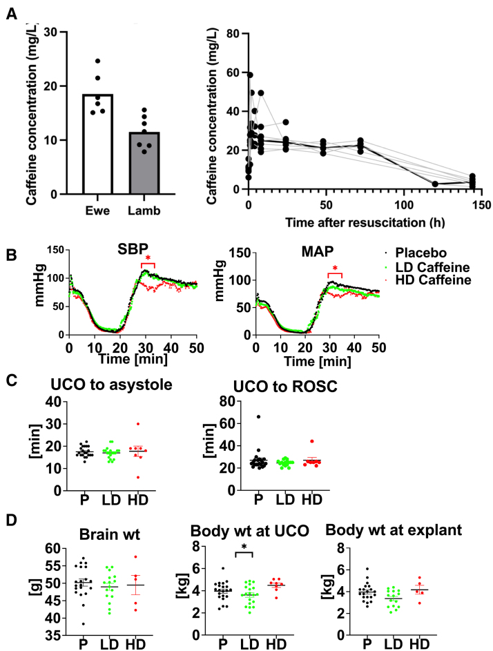
Methods: Ewes were randomly assigned to receive either 1 g IV caffeine citrate or placebo before delivery and placental transport assessed. Near-term lambs (141-143 days) of both sexes were subjected to severe global hypoxia-ischemia utilizing an acute umbilical cord occlusion model. Lambs that received caffeine in utero also received 20 mg/kg IV caffeine citrate following resuscitation and 10 mg/(kg·d) IV for 2 days. An additional cohort received 60 mg/kg followed by 30 mg/(kg·d) (low dose versus high dose) postnatally. Biochemical, histological, and neurological outcome measures in lambs were assessed over a 6-day period.

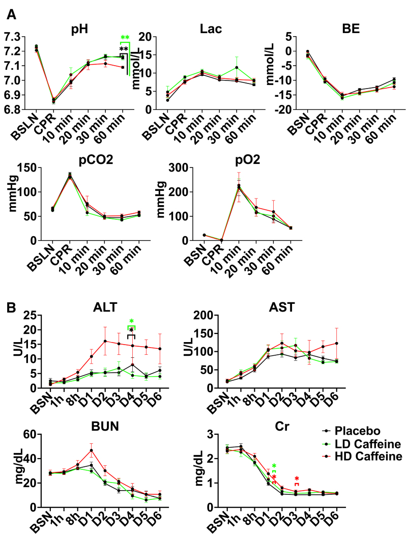
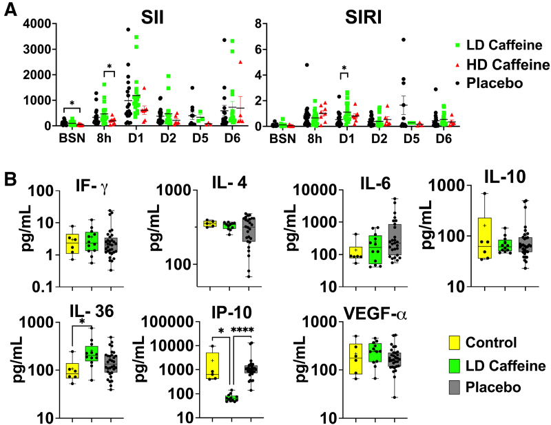
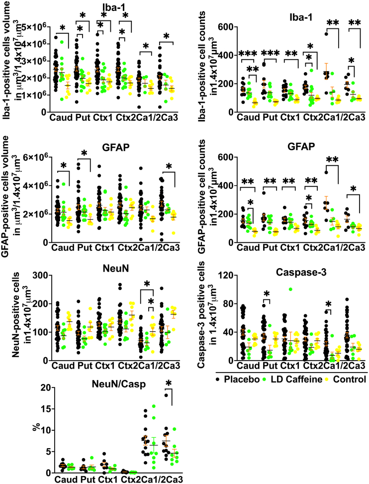
Results: Perinatal caffeine administration demonstrated excellent placental transport kinetics and was well tolerated with lamb plasma levels comparable to those targeted in neonates with apnea of prematurity. Caffeine administration resulted in a systemic immunomodulatory effect, evidenced by significant reductions in proinflammatory IP-10 levels. Treated lambs demonstrated improved neurodevelopmental outcomes, while histological analysis revealed that caffeine reduced gray matter injury and attenuated inflammation in the cingulate and parasagittal cortex. This neuroprotective effect was greater and via a different mode of action than we previously reported for azithromycin. A higher caffeine dosing regimen demonstrated significant toxicity.

Conclusions: Perinatal caffeine administration is well tolerated, attenuates systemic and brain inflammation, and contributes to improvements in histological and neurological outcomes in an ovine model of neonatal hypoxic-ischemic encephalopathy.

Keywords: caffeine; developing countries; gray matter; hypoxia-ischemia, brain; sheep.


Conflict of interest statement

None.

Figures

Figure 1.
Figure 1.
Caffeine pharmacokinetics and effects on resuscitation. A, Caffeine concentration in ewe (n=6) and lamb (n=7) plasma at the time of umbilical cord occlusion and caffeine concentration in lamb (n=7) plasma over time. B, Hemodynamic changes in response to umbilical cord occlusion (UCO) with drop in systolic blood pressure (SBP) and mean arterial pressure (MAP) followed by increase reflecting return of spontaneous circulation (ROSC). Hemodynamic data were analyzed using grouped analysis of the individual group’s means for a specific time point. Placebo (P): n=19; low-dose (LD) caffeine: n=14; and high-dose (HD) caffeine: n=7. C, Time to asystole and time to ROSC were similar among the compared groups. Groups were compared using the Kruskal-Wallis test. Placebo: n=21; LD caffeine: n=18–19; and HD caffeine: n=8. D, Selected anthropometric parameters between the studied groups. Brain and body weight differences were assessed using ANOVA. Data in graph B are shown as mean and graph C and D as mean±SEM. Placebo: n=20–21; LD caffeine: n=16–19; and HD caffeine: n=5–8. LD-caffeine–treated group is presented in green, HD-caffeine–treated group is presented in red, and placebo is presented in black. *P<0.05. wt indicates weight.
Figure 2.
Figure 2.
Biochemical parameters in treated vs placebo groups. A, Umbilical cord occlusion (UCO) leads to profound acidosis in all groups. High-dose (HD)-caffeine–treated lambs demonstrated greater acidosis at 60 minutes compared with the low-dose (LD)-caffeine and placebo groups. All studied groups exhibited similar hyperlactatemia, base excess, carbon dioxide, and oxygen levels. B, HD caffeine demonstrated significant toxicity evidenced by elevated alanine transaminase (ALT) levels on day 5, as well as creatinine on days 2 and 4. No differences in aspartate aminotransferase (AST) and blood urea nitrogen (BUN) levels were noted between the studied groups. The data were analyzed using mixed-effects analysis with Tukey correction for multiple comparisons. Data in the graphs represent mean±SEM. For A, HD caffeine, n=5–8; LD caffeine, n=19–20; and placebo, n=21. For B, HD caffeine, n=4–5; LD caffeine, n=5–15; and placebo, n=7–20. LD-caffeine–treated group is presented in green, HD-caffeine–treated group is presented in red, and placebo is presented in black. *P<0.05, **P<0.01. BE indicates base excess; BSN/BSLN, pre-UCO baseline; CPR, cardiopulmonary resuscitation; and Lac, lactate.
Figure 3.
Figure 3.
Peripheral markers of inflammation. A, The peripheral blood cell index systemic immune-inflammation index (SII=ANC×PLT/ALC) was suppressed in high-dose (HD) caffeine at baseline (BSN) compared with placebo, and at 8 hours after umbilical cord occlusion (UCO) compared with low-dose (LD)-caffeine group. Systemic inflammation response index (SIRI=ANC×Mono/ALC) was elevated in the LD-caffeine group compared with the placebo on day 1. The SII and SIRI scores were evaluated by mixed-effect analysis with Tukey correction for multiple comparisons. The summary column graphs are showing means±SEM. HD caffeine: n=3–8; LD caffeine: n=3–19, and placebo: n=10–21. B, At 6 days after the UCO, we measured changes in IP-10 in the LD-caffeine group compared with the placebo, as well as in age-matched control and in IL-36 that was higher in the LD-caffeine group compared with control. No changes were observed in IL (interleukin)-4, IL-6, IF (interferon)-γ, IP (interferon gamma-induced protein 10)-10, or VEGF (vascular endothelial growth factor A)-α. We used Mann-Whitney U test. LD caffeine: n=11–12; and placebo: n=26–32. The box graphs show mean by plus sign and median with interquartile range. *P<0.05, ****P<0.0001. LD-caffeine–treated group is presented in green, control is presented in yellow, and placebo is presented in black. ALC indicates absolute lymphocyte count; ANC, absolute neutrophil count; Mono, monocytes; and PLT, platelets.
Figure 4.
Figure 4.
Histological changes in gray matter. We compared quantitative changes in inflammatory markers of gliosis (GFAP [glial fibrillary acidic protein]), microglial accumulation (Iba-1 [ionized calcium-binding adapter molecule 1]), neuronal counts (NeuN), cellular death markers (Casp-3) in cingulate gyrus Ctx (Ctx1), first parasagittal gyrus Ctx (Ctx2), caudate (Caud), putamen (Put), and Ca1/2 (Ca1/2) and Ca3 (Ca3) of the hippocampus. Placebo animals (n=33–41) were compared with the low-dose (LD)-caffeine–treated animals (n=7–11) and controls (n=4–9) using ANOVA or Kruskal-Wallis test as appropriate. Data are presented as mean±SEM. Brackets show significance as follows: *P<0.05. LD-caffeine–treated group is presented in green, control is presented in yellow, and placebo is presented in black.
Figure 5.
Figure 5.
Quantitative analysis of white matter markers and markers of inflammation: while overall white matter structure was not significantly altered by umbilical cord occlusion (UCO), there is more cell death in the placebo group compared with low-dose (LD) caffeine, reflected by a higher number of cells labeled with cleaved caspase-3. More inflammation was noted in the SCWM2 in the placebo group, reflected by the accumulation of microglia and higher microglial volumes. Placebo lamb histologies (n=31–41) were compared with the LD-caffeine–treated animals (n=7–12) and controls (n=5–9) using ANOVA or Kruskal-Wallis test as appropriate. Data are presented as mean±SEM. Brackets show significance as follows: *P<0.05. LD-caffeine–treated group is presented in green, control is presented in yellow, and placebo is presented in black. CC-1 indicates adenomatous polyposis coli protein; GFAP, glial fibrillary acidic protein; Iba-1, ionized calcium binding adaptor molecule 1; MBP, myelin basic protein; PVWM, periventricular white matter; and SCWM, subcortical white matter.
Figure 6.
Figure 6.
Neurodevelopmental outcomes following caffeine or azithromycin treatment. A, We assessed composite and individual outcomes consisting of motor function, feeding, and activity and assigned a severity score. The summary graphs picture the relationship of low-dose (LD) and high-dose (HD) caffeine to placebo animals. LD-caffeine–treated group is presented in blue (n=19), HD-caffeine–treated group is presented in red (n=8), placebo is presented in black (n=53), azithromycin is presented in green (n=21), and control is presented in purple (n=12). B, The correlation matrix depicts Spearman correlation coefficients of selected study parameters with combined neurological outcomes scores on day 6 after umbilical cord occlusion (UCO) that reached statistical significance (P<0.05). ALT, alanine transaminase; ANC, absolute neutrophil count; AST, aspartate aminotransferase; BUN, blood urea nitrogen; Mono, monocytes; SII, systemic immune-inflammation index; SIRI, system inflammation response index; and wt, weight.

References

    1. Olusanya BO, Nair MKC, Wright SM, Hadders-Algra M; Global Research on Developmental Disabilities Collaborators (GRDDC). Global progress towards early childhood development for children with disabilities, 2013-2023. BMJ Paediatr Open. 2023;7:e002046. doi: 10.1136/bmjpo-2023-002046 - PMC - PubMed
    1. Mike JK, Wu KY, White Y, Pathipati P, Ndjamen B, Hutchings RS, Losser C, Vento C, Arellano K, Vanhatalo O, et al. . Defining longer-term outcomes in an ovine model of moderate perinatal hypoxia-ischemia. Dev Neurosci. 2022;44:277–294. doi: 10.1159/000525150 - PMC - PubMed
    1. Thayyil S, Pant S, Montaldo P, Shukla D, Oliveira V, Ivain P, Bassett P, Swamy R, Mendoza J, Moreno-Morales M, et al. ; HELIX Consortium. Hypothermia for moderate or severe neonatal encephalopathy in low-income and middle-income countries (HELIX): a randomised controlled trial in India, Sri Lanka, and Bangladesh. Lancet Glob Health. 2021;9:e1273–e1285. doi: 10.1016/S2214-109X(21)00264-3 - PMC - PubMed
    1. Ranjan AK, Gulati A. Advances in therapies to treat neonatal hypoxic-ischemic encephalopathy. J Clin Med. 2023;12:6653. doi: 10.3390/jcm12206653 - PMC - PubMed
    1. Victor S, Rocha-Ferreira E, Rahim A, Hagberg H, Edwards D. New possibilities for neuroprotection in neonatal hypoxic-ischemic encephalopathy. Eur J Pediatr. 2022;181:875–887. doi: 10.1007/s00431-021-04320-8 - PMC - PubMed

Publication types