Review
. 2024 Sep 19;16(9):e69691.
doi: 10.7759/cureus.69691. eCollection 2024 Sep.

Cutaneous Basal Cell Carcinoma In Situ: A Review of the World Literature

Affiliations
Review

Cutaneous Basal Cell Carcinoma In Situ: A Review of the World Literature

Philip R Cohen et al. Cureus. .

Abstract

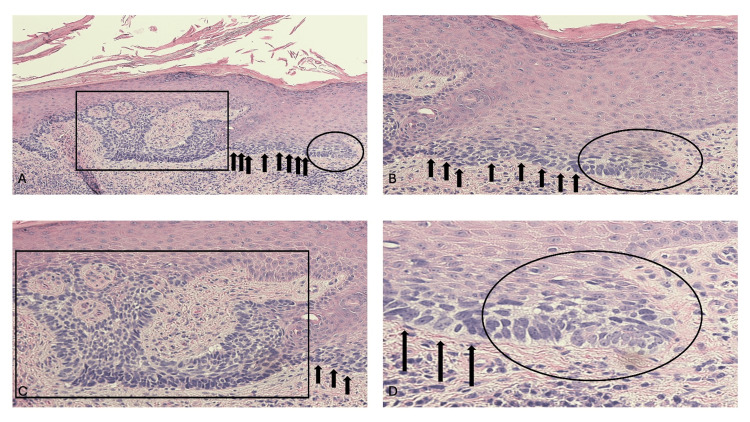
Cutaneous basal cell carcinoma (BCC) in situ is a recently recognized subtype of the skin neoplasm in which the abnormal cells are confined to the epidermis. BCC in situ of the skin was previously referred to as a superficial BCC. A review of the world literature has revealed 10 cutaneous BCCs in situ that have been described in nine patients but likely reflect a more general phenomenon. The neoplasm typically presents as an asymptomatic red plaque on the abdomen, upper extremity, back, and chest. Pathologic changes frequently show confluent tumor cells along the epidermal basal layer or superficial aggregates of neoplastic cells that are contiguous with the epidermis and extend into the dermis. Genomic evaluation has been performed in neoplasms from one individual with cutaneous BCC in situ and metastatic BCC; like other variants of BCC, an aberration of the PTCH1 gene was observed. In contrast to his liver metastasis, the in situ carcinoma had a lower tumor mutational burden, lacked programmed death-ligand 1 (PD-L1) and programmed death-ligand 2 (PD-L2) amplification and had a distinct PTCH1 mutation, suggesting that the in situ BCC of his skin and the metastatic BCC of his liver were derived from different clones of cells.

Keywords: basal; cancer; carcinoma; cell; cutaneous; fibroepithelioma; in situ; invasive; molecular; skin.


Conflict of interest statement

Conflicts of interest: In compliance with the ICMJE uniform disclosure form, all authors declare the following: Payment/services info: All authors have declared that no financial support was received from any organization for the submitted work. Financial relationships: Razelle Kurzrock declare(s) personal fees from Genentech, Merck Serono, Pfizer, Boehringer Ingelheim, TopAlliance, Takeda, Incyte, Debiopharm, Medimmune, Sequenom, Foundation Medicine, Konica Minolta, Grifols, Omniseq, and Guardant. Razelle Kurzrock has received research funding from these companies. Razelle Kurzrock declare(s) personal fees from X-Biotech, Caris, Datar Cancer Genomics, Neomed, Pfizer, Actuate Therapeutics, and Roche, . Razelle Kurzrock has been a consultant and/or speaker fees and/or advisory board of these companies. Razelle Kurzrock declare(s) non-financial support from IDbyDNA and CureMatch Inc. Razelle Kurzrock has an equity interest in these companies. Razelle Kurzrock declare(s) non-financial support from CureMatch and CureMetrix. Razelle Kurzrock serves on the Board of CureMatch and CureMetrix, and is a co-founder of CureMatch. Other relationships: All authors have declared that there are no other relationships or activities that could appear to have influenced the submitted work.

Figures

Figure 1
Figure 1. Clinical presentation of a basal cell carcinoma (BCC) in situ of the skin on the right dorsal hand
A 51-year-old male with a history of actinic keratoses and basal cell carcinomas (BCCs) presented with a painless, non-pruritic lesion that had been present for less than a year and had progressively increased in size. The BCC in situ appeared as 2 x 1-centimeter scaly erythematous plaques with a raised peripheral edge on the right dorsal hand proximal to the thumb (black oval); a shave biopsy was performed. The two erythematous patches (black arrows) are the sites of actinic keratoses that were treated with liquid nitrogen cryotherapy. In addition, features of vitiligo are demonstrated by the depigmentation of the distal dorsal hand and the fingers.
Figure 2
Figure 2. Microscopic features of a cutaneous basal cell carcinoma (BCC) in situ on the right dorsal hand of a 51-year-old male
Distant (A) and closer (B, C, and D) views demonstrate the pathologic changes the right dorsal hand BCC in situ. There is compact orthokeratosis (demonstrated by thickening of the keratin layer in the stratum corneum with preserved keratinocyte maturation and without retained nuclei) overlying the epidermis (which is the outermost layer of the skin which is composed of several strata from the most superficial to the deepest including the stratum corneum, stratum lucidum, stratum granulosum, stratum spinosum, and stratum basale); this finding correlates with the scaling observed clinically. Superficial buds of basaloid tumor cells that are contiguous with the epidermis extend into the papillary dermis and anastomose (shown within the black rectangle); also, there is an aggregate of tumor cells present in the lower layers of the epidermis (within the oval). At the periphery of the anastomosing aggregates of in situ BCC, there is peripheral palisading of the tumor keratinocytes. In addition, there is a confluent proliferation of atypical cells along the basal layer of the epidermis (black arrows). Importantly, there were no basaloid tumor cells that were not contiguous with the epidermis, in the dermis. In the papillary dermis there is a lymphocytic inflammatory infiltrate that is predominantly around the blood vessels, but also diffusely present between the collagen bundles. Correlation of the clinical morphology and the pathologic findings established a diagnosis of cutaneous BCC in situ. The residual tumor was excised and there was no recurrence of the in situ carcinoma after three-and-a-half years (hematoxylin and eosin: A, x10; B, x20; C, x 20; D, x 40).

Similar articles

References

    1. Basal cell carcinoma: comprehensive clinical and histopathological aspects, novel imaging tools and therapeutic approaches (review) Niculet E, Craescu M, Rebegea L, et al. Exp Ther Med. 2022;23:60. - PMC - PubMed
    1. Basal cell carcinoma: an updated review of pathogenesis and treatment options. Hernandez LE, Mohsin N, Levin N, Dreyfuss I, Frech F, Nouri K. Dermatol Ther. 2022;35:0. - PubMed
    1. Basal cell carcinoma: a narrative review on contemporary diagnosis and management. Naik PP, Desai MB. Oncol Ther. 2022;10:317–335. - PMC - PubMed
    1. Advances in management and therapeutics of cutaneous basal cell carcinoma. Chen OM, Kim K, Steele C, et al. Cancers (Basel) 2022;14:14–16. - PMC - PubMed
    1. Basal cell carcinoma. Heath MS, Bar A. Dermatol Clin. 2023;41:13–21. - PubMed

LinkOut - more resources