Healing of a Large Periapical Lesion Using Non-Surgical Root Canal Retreatment: A Case Report
- PMID: 39429432
- PMCID: PMC11486631
- DOI: 10.7759/cureus.69586
Healing of a Large Periapical Lesion Using Non-Surgical Root Canal Retreatment: A Case Report
Abstract
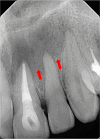
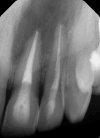
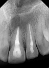
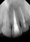
This case report highlights the successful healing of a large periapical lesion through non-surgical root canal retreatment. A 29-year-old male patient presented with a significant radiolucency associated with teeth #21 and #22, initially treated non-surgically. Despite the lesion's size, the treatment, which included thorough canal disinfection and obturation, led to substantial healing. A follow-up cone-beam computed tomography (CBCT) scan after one year confirmed the buccal cortical bone reformation and improvement in the incisive canal area except for the apical region of #21. Subsequently, root canal retreatment was performed for #21. Complete healing was achieved after two years, demonstrating that even extensive periapical lesions can be effectively treated with non-surgical endodontic retreatment, avoiding invasive surgical intervention.
Keywords: cone-beam computed tomography (cbct); effective disinfectant protocol; large periapical lesion; non-surgical endodontic treatment; single-visit endodontic treatment; teeth requiring non-surgical endodontic treatment.
Copyright © 2024, Alyousef et al.
Conflict of interest statement
Human subjects: Consent was obtained or waived by all participants in this study. Prince Sultan Military Medical City IRB committee issued approval PSMMC #1851793. Conflicts of interest: In compliance with the ICMJE uniform disclosure form, all authors declare the following: Payment/services info: All authors have declared that no financial support was received from any organization for the submitted work. Financial relationships: All authors have declared that they have no financial relationships at present or within the previous three years with any organizations that might have an interest in the submitted work. Other relationships: All authors have declared that there are no other relationships or activities that could appear to have influenced the submitted work.
Figures


References
-
- Taxonomy, ecology, and pathogenicity of the root canal flora. Sundqvist G. Oral Surg Oral Med Oral Pathol. 1994;78:522–530. - PubMed
-
- Oral surgery—oral pathology conference no. 17, Walter Reed Army Medical Center: periapical lesions—types, incidence, and clinical features. Bhaskar SN. Oral Surg Oral Med Oral Pathol. 1966;21:657–671. - PubMed
-
- Evaluation of pathologists (histopathology) and radiologists (cone beam computed tomography) differentiating radicular cysts from granulomas. Rosenberg PA, Frisbie J, Lee J, et al. J Endod. 2010;36:423–428. - PubMed
-
- Cleaning and shaping the root canal. Schilder H. Dent Clin North Am. 1974;18:269–296. - PubMed
Publication types
LinkOut - more resources
Full Text Sources
