. 2024 Sep 16;32(4):101342.
doi: 10.1016/j.omtm.2024.101342. eCollection 2024 Dec 12.

A novel AAV9-dual microRNA-vector targeting GRIK2 in the hippocampus as a treatment for mesial temporal lobe epilepsy

Affiliations

A novel AAV9-dual microRNA-vector targeting GRIK2 in the hippocampus as a treatment for mesial temporal lobe epilepsy

Stéphane J Baudouin et al. Mol Ther Methods Clin Dev. .

Abstract

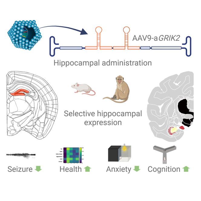
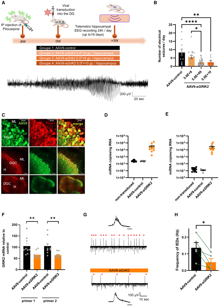
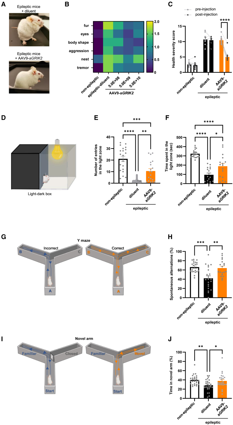
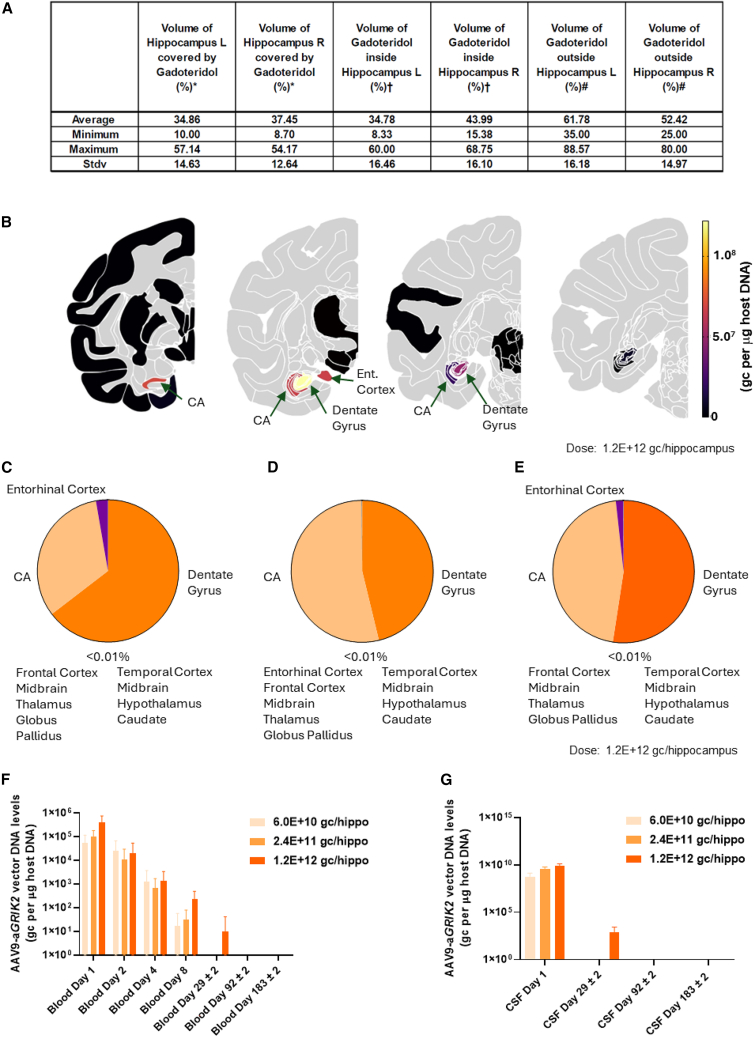
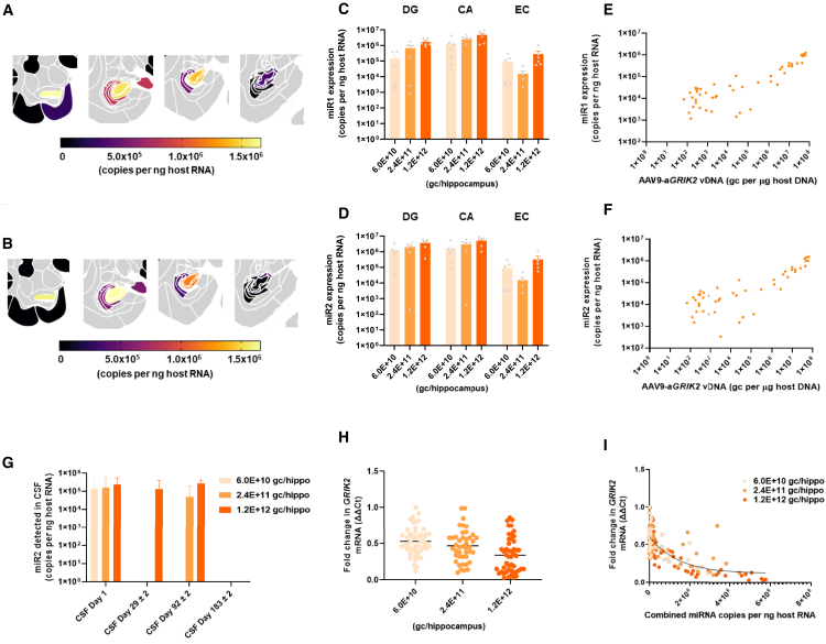
Mesial temporal lobe epilepsy (mTLE) is the most prevalent type of epilepsy in adults. First and subsequent generations of anti-epileptic therapy regimens fail to decrease seizures in a large number of patients suffering from mTLE, leaving surgical ablation of part of the hippocampus as the only therapeutic option to potentially reach seizure freedom. GluK2 has recently been identified as a promising target for the treatment of mTLE using gene therapy. Here, we engineered an adeno-associated virus serotype 9 vector expressing a cluster of two synthetic microRNAs (miRNAs), expressed from the human synapsin promoter, that target GRIK2 mRNA. Intra-hippocampal delivery of this vector in a mouse model of mTLE significantly reduced GRIK2 expression and daily seizure frequency. This treatment also improved the animals' health, reduced their anxiety, and restored working memory. Focal administration of the vector to the hippocampus of cynomolgus monkeys in GLP toxicology studies led to the selective transduction of hippocampal neurons with little exposure elsewhere in the brain and no transduction outside the central nervous system. Expression of miRNAs in hippocampal neurons resulted in substantially decreased GRIK2 mRNA expression. These data suggest that the intra-hippocampal delivery of a GMP-grade AAV9 encoding a synthetic miRNAs targeting GRIK2 is a promising treatment strategy for mTLE.

Keywords: AAV9; GRIK2; epilepsy; gene therapy; microRNA.


Conflict of interest statement

A patent application has been filed relating to this work. S.J.B., N.Pearson, C.H., N.Partouche, J.G., and R.P. declare an association with uniQure/Corlieve Therapeutics. M.W. and I.B. declare an association with uniQure B.V. A.G., J.S., A.M., and O.D. declare association with Regenxbio Inc.

Figures

None
Graphical abstract
Figure 1
Figure 1
Design, selection, and optimization of synthetic miRNA constructs capable of efficient GluK2 knockdown with favorable miRNA processing profiles (A) Schematics depicting the vector genome formats of Gen1.1-.3 and Gen2.1-.2 constructs. (B) Quantification of firefly luciferase(ffluc)-GluK2 reporter knockdown mediated by siRNA and dual reporter plasmid transfection in 293 cells. Data are reported as ffluc RLU from co-transfection of experimental siRNAs with ffluc-GluK2 reporter relative to ffluc RLU from co-transfection of negative control siRNA with ffluc-GluK2 reporter, after normalization to control renilla luciferase RLU (reported as mean ± SEM). (C) Quantification of GRIK2 mRNA levels following miRNA plasmid construct transfection of Gen1.1 constructs into iCell GlutaNeurons, relative to lipid only transfection control (NT), as measured by real-time qPCR and reported as mean ± SD. (D) Quantification of relative levels of mature guide and passenger strand expression from top GRIK2-targeting Gen1.1 constructs as measured by smRNA-seq following plasmid transfection of iCell GlutaNeurons. (E) Assessment of the impact of optimization efforts on G:P by smRNA-seq following Gen1.1 and Gen1.2 plasmid transfection of N2A cells. (F) Assessment of the impact of miRNA concatemerization and addition of a stuffer sequence on G:P by smRNA-seq following plasmid transfection of N2A cells. (G) Quantification of GRIK2 mRNA levels by real-time qPCR (normalized to GAPDH) following transduction of C57Bl6/J mouse cortical neurons with AAV9 vectors packaging Gen1.3, Gen2.1, and Gen2.2 constructs; data are reported as mean ± SEM, NT = non-transduced. (H) Quantification of GluK2 protein levels by western blot (normalized to β-actin) following transduction of C57Bl6/J mouse cortical neurons with AAV9 vectors packaging Gen1.3, Gen2.1, and Gen2.2 constructs. ∗∗p < 0.01, ∗∗∗∗p < 0.0001 one-way ANOVA, followed by Dunnett’s multiple comparison. Data are reported as mean ± SEM, NT = non-transduced.
Figure 2
Figure 2
Administration of AAV9-aGRIK2 decreases GluK2 protein expression in hippocampal neurons in vivo (A) Schematic showing the two hippocampal injection sites for diluent, AAV9-control (5.0E+9 gc/hippocampus), or AAV9-aGRIK (at different doses) (orange); the corresponding in situ hybridization for miR1, at the sites of injection, is on the right. (B) Quantification of the vector genome copies in the hippocampus. (C) Quantification of miR1 in the hippocampus. (D) Quantification of miR2 in the hippocampus. (E) Correlation between vector copies and miRNA expression. (F) GluK2 protein expression up to 6 months after administration of AAV9-aGRIK2 or diluent in pilocarpine-treated epileptic mice. ∗∗p < 0.01, ∗∗∗∗p < 0.0001 one-way ANOVA, followed by Dunnett’s multiple comparison. Data are reported as mean ± SEM.
Figure 3
Figure 3
AAV9-aGRIK2 treatment decreases seizure in epileptic mice and inter-ictal epileptiform discharges in human organotypic hippocampal slices (A) Timeline of the experimental procedure in epileptic mice (mTLE, top) injected bilaterally in the hippocampus with either AAV9-control (5.0E+9 gc/hippocampus) or AAV9-GRIK2 (at different doses) and example of a typical electrographic seizure (ictal event, bottom). (B) Number of seizures per day in the presence of either AAV9-control or AAV9-GRIK2. ∗p < 0.05, ∗∗p < 0.01, and ∗∗∗∗p < 0.0001, one-way ANOVA followed by Tukey’s multiple comparisons test. (C) Photomicrographs showing a representative example of dentate gyrus granule cells (DGCs) transduced with AAV9-GFP (AAV9.hSyn1.GFP) in hippocampal organotypic slices from a patient with temporal lobe epilepsy (TLE), using double immunostaining with anti-Prox1 (left), and anti-GFP (middle) antibodies; Molecular layer (ML), hilus (H), scale bars, top, 50 μm, middle, 100 μm, and bottom, 200 μm. (D) Quantification of miR1 expression level. (E) Quantification of mi2R expression level. (F) Quantification of GRIK2 mRNA in tissue treated with AAV9-aGRIK2 normalized to treatment with the AAV9-control. Student’s t test, ∗∗p < 0.01. (G) Representative traces of LFPs recorded in human hippocampal organotypic slices transduced with either control AAV9-control (top) or AAV9-aGRIK2 (bottom, 2.0E+10 gc per slices); enlarged interictal-like epileptiform discharges (IEDs) shown as upper and lower traces (scale bar, 50 ms); note the decrease of IEDs in slices treated with AAV9-aGRIK2. (H) Quantification of the frequency of IEDs in human organotypic hippocampal slices from five patients with mTLE treated with either AAV9-control or AAV9-aGRIK2; control and treated slices from the same patient are paired (lines) for Student’s t test, ∗p < 0.05. Data are reported as mean ± SEM.
Figure 4
Figure 4
AAV9-aGRIK2 treatment improves comorbidities in epileptic mice (A) Representative images of pilocarpine mice treated with either diluent (top) or AAV9-aGRIK2 (bottom). (B) Health severity score categories measured in naive non-epileptic and epileptic mice treated with either diluent or AAV9-aGRIK2; yellow is low health and dark blue normal health. (C) Health severity score of naive non-epileptic and epileptic mice with either diluent or AAV9-aGRIK2. ∗∗∗∗p < 0.0001, two-way ANOVA repeated measure followed by Sidak’s multiple comparison test. (D) Schematic representation of the light-dark box. (E) Numbers of entry into the light zone. (F) Time spent in the light zone. (G) Schematic representation of the Y-Maze. (H) Spontaneous correct alternation in the Y-Maze (%). (I) Schematic representation of the novel arm maze. (J) Time spent in the novel arm (%). ∗p < 0.05, ∗∗p < 0.01, ∗∗∗p < 0.001, ∗∗∗∗p < 0.0001, one-way ANOVA Tukey’s multiple comparisons test in (E)–(J). Data are reported as mean ± SEM.
Figure 5
Figure 5
Expression of AAV9-aGRIK2 vector DNA is restricted to the hippocampus in non-human primates (A) Volume of distribution of gadoteridol inside and outside of the hippocampus. (B) Expression level of the vector DNA in tissue punches sampled in relevant brain regions, after intra-hippocampal administration of AAV9-aGRIK2 at 1.2E+12 gc/hippocampus (black: below detection level and gray: not evaluated, CA: cornu ammonis). (C) Expression of the vector DNA in punches from different brain regions (expressed as percentage of the total amount of vector DNA quantified) after administration AAV9-aGRIK2 at 6.0E+10 gc/hippocampus, (D) 2.4E+11 gc/hippocampus and (E) 1.2E+12 gc/hippocampus. (F) Expression of the vector DNA in the blood serum and (G) in the CSF after administration of AAV9-aGRIK2 at 6.0E+10 gc/hippocampus, 2.4E+11 gc/hippocampus and 1.2E+12 gc/hippocampus. Data are reported as mean ± SEM.
Figure 6
Figure 6
Knockdown of GRIK2 following administration of AAV9-aGRIK2 is restricted to the hippocampus (A) Expression level of miR1 and (B) miR2 after intra-hippocampal administration of AAV9-aGRIK2 at 1.2E+12 gc/hippocampus. (C) Expression levels of miR1 and (D) miR2 in different brain regions after administration AAV9-aGRIK2 at 6.0E+10 gc/hippocampus, 2.4E+11 gc/hippocampus and 1.2E+12 gc/hippocampus. (E) Correlation between vector copies and miR1 or (F) miR2 expression. (G) Detection of miR2 in the CSF after administration of AAV9-aGRIK2 at 6.0E+10 gc/hippocampus, 2.4E+11 gc/hippocampus and 1.2E+12 gc/hippocampus. (H) Expression levels of GRIK2 mRNA in the hippocampus and EC after administration AAV9-aGRIK2 at 6.0E+10 gc/hippocampus, 2.4E+11 gc/hippocampus, and 1.2E+12 gc/hippocampus compared with the diluent. (I) Correlation between combined expression of the two miRNAs and the vector DNA. Data are reported as mean ± SEM.

References

    1. Cross J.H. Epilepsy in the WHO European region: Fostering epilepsy care in Europe. Epilepsia. 2011;52:187–188. doi: 10.1111/J.1528-1167.2010.02903.X. - DOI - PubMed
    1. Semah F., Picot M.C., Adam C., Broglin D., Arzimanoglou A., Bazin B., Cavalcanti D., Baulac M. Is the underlying cause of epilepsy a major prognostic factor for recurrence? Neurology. 1998;51:1256–1262. doi: 10.1212/WNL.51.5.1256. - DOI - PubMed
    1. Asadi-Pooya A.A., Stewart G.R., Abrams D.J., Sharan A. Prevalence and Incidence of Drug-Resistant Mesial Temporal Lobe Epilepsy in the United States. World Neurosurg. 2017;99:662–666. doi: 10.1016/J.WNEU.2016.12.074. - DOI - PubMed
    1. Phuong T.H., Houot M., Méré M., Denos M., Samson S., Dupont S. Cognitive impairment in temporal lobe epilepsy: contributions of lesion, localization and lateralization. J. Neurol. 2021;268:1443–1452. doi: 10.1007/s00415-020-10307-6. - DOI - PubMed
    1. Tai X.Y., Bernhardt B., Thom M., Thompson P., Baxendale S., Koepp M., Bernasconi N. Blackwell Publishing Ltd; 2018. Review: Neurodegenerative Processes in Temporal Lobe Epilepsy with Hippocampal Sclerosis: Clinical, Pathological and Neuroimaging Evidence. - DOI - PubMed

LinkOut - more resources