

A Review on Low-Dose Emission Tomography Post-Reconstruction Denoising with Neural Network Approaches
- PMID: 39429805
- PMCID: PMC11486494
- DOI: 10.1109/trpms.2023.3349194
A Review on Low-Dose Emission Tomography Post-Reconstruction Denoising with Neural Network Approaches
Abstract
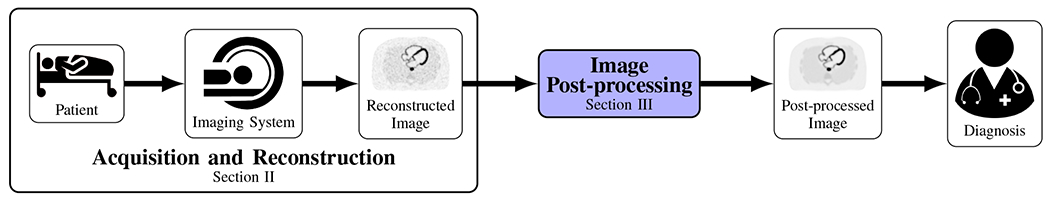
Low-dose emission tomography (ET) plays a crucial role in medical imaging, enabling the acquisition of functional information for various biological processes while minimizing the patient dose. However, the inherent randomness in the photon counting process is a source of noise which is amplified low-dose ET. This review article provides an overview of existing post-processing techniques, with an emphasis on deep neural network (NN) approaches. Furthermore, we explore future directions in the field of NN-based low-dose ET. This comprehensive examination sheds light on the potential of deep learning in enhancing the quality and resolution of low-dose ET images, ultimately advancing the field of medical imaging.
Keywords: Deep Learning; Low-Dose; PET; SPECT.
Figures


References
-
- Mattsson S and Söderberg M, “Radiation dose management in CT, SPECT/CT and PET/CT techniques,” Radiation protection dosimetry, vol. 147, no. 1-2, pp. 13–21, 2011. - PubMed
-
- Gimelli A, Achenbach S, Buechel RR, Edvardsen T, Francone M, Gaemperli O, Hacker M, Hyafil F, Kaufmann PA, Lancellotti P, et al. “Strategies for radiation dose reduction in nuclear cardiology and cardiac computed tomography imaging: A report from the European Association of Cardiovascular Imaging (EACVI), the Cardiovascular Committee of European Association of Nuclear Medicine (EANM), and the European Society of Cardiovascular Radiology (ESCR),” European heart journal, vol. 39, no. 4, pp. 286–296, 2018. - PubMed
-
- Anger HO, “Scintillation camera,” Review of scientific instruments, vol. 29, no. 1, pp. 27–33, 1958.
-
- Natterer F, The Mathematics of Computerized Tomography (Classics in Applied Mathematics). SIAM, 2001.
-
- Shepp LA and Vardi Y, “Maximum likelihood reconstruction for emission tomography,” IEEE transactions on medical imaging, vol. 1, no. 2, pp. 113–122, 1982 - PubMed
Grants and funding
LinkOut - more resources
Full Text Sources