Review
. 2024 Jun 6;9(9):417-424.
doi: 10.1016/j.vgie.2024.05.015. eCollection 2024 Sep.

EUS-guided hepaticogastrostomy: practical tips and tricks

Affiliations
Review

EUS-guided hepaticogastrostomy: practical tips and tricks

Kambiz Kadkhodayan et al. VideoGIE. .

Abstract

Background and aims: EUS-guided hepaticogastrostomy (EUS-HGS) has gained traction as a reliable and safe method for definitive biliary drainage in patients who cannot undergo traditional transampullary procedures. Many of the newly developed single-stage devices are not yet approved for clinical use in Western practice.

Methods: We highlight key clinical and technical aspects of EUS-HGS using devices that are currently available in Western countries. The article is a comprehensive step-by-step technical review of EUS-HGS, and the video demonstrates high-level tips to overcome commonly encountered procedural challenges.

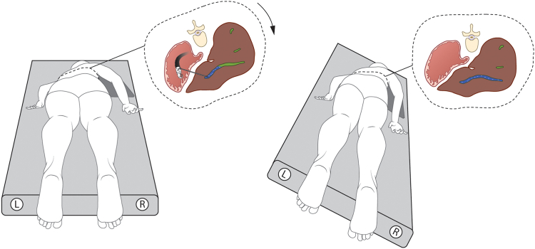
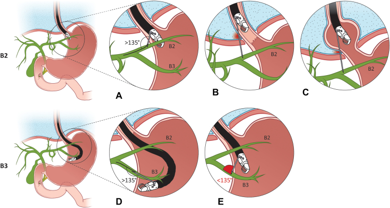
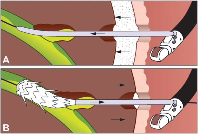
Results: Patients with biliary obstruction underwent EUS-HGS at our center. The technical difficulties that were encountered are highlighted in the article and the accompanying video. The article and video provide a detailed review of (1) preprocedural considerations, (2) puncture site selection, (3) biliary puncture, (4) contrast injection, (5) guidewire manipulation, (6) tract dilation, and (7) stent placement.

Conclusion: An understanding and implementation of the technical nuances highlighted in this article should help Western endoscopists navigate the complexities of EUS-HGS and ensure optimal outcomes.


Conflict of interest statement

Dr Irani is a consultant for Boston Scientific, WL Gore & Associates. The other author disclosed no financial relationships relevant to this publication.

Figures

Figure 1
Figure 1
Modified prone position. Note that the right side of the patient is lowered by 10° to 20°. This lowers the antidependent (right-side) intrahepatic ducts. In this position, gravity-related pooling of contrast favors a more diffuse distribution within the biliary tree.
Figure 2
Figure 2
Key differences between EUS-guided access when using segment 2 (B2) and segment 3 (B3) intrahepatic ducts. (A) Ideal puncture site for B2 ducts located 2 to 3 cm below the squamocolumnar junction (SQJ). (B) Risk of transpleuritic access if the puncture site is above the SQJ. (C) Patient with a hiatal hernia. Note that the SQJ is located within the chest cavity, and despite puncturing 2 to 3 cm below the SQJ, the pleura is breached. (D) Optimal endoscope position and puncture site to access B3. The endoscope is in a deep (caudal) and flexed position. Note the favorable (>135°) angle of approach. (E) Suboptimal endoscope position and puncture site for B3 access. The endoscope is in a shallow (cranial) and straight position. Note the unfavorable (<135°) angle of approach.
Figure 3
Figure 3
Optimal parameter when choosing an intrahepatic duct for puncture.
Figure 4
Figure 4
EUS image of the gastrohepatic ligament.
Figure 5
Figure 5
(A) Stent placement across the gastrohepatic ligament. (B) Position-dependent kinking of the stent that may occur as the stomach is pulled away from the undersurface of the liver.
Figure 6
Figure 6
Optimal angle of approach between the needle and targeted bile duct. (a) Longitudinal axis of needle and guidewire. (b) Longitudinal axis of bile duct.
Figure 7
Figure 7
Different layers that comprise the hepaticogastrostomy tract.
Figure 8
Figure 8
Traditional (left) and segmental (right) tract dilation method. (A) Biliary puncture and guidewire insertion. (B) Balloon dilation. Note the separate dilation of the bile duct side (1) and gastric side (2) when using the segmental dilation method. (C) Post-dilation bile leak (left). Note the absence of bile leak (right) due to the non-dilated segment of liver parenchyma.
Figure 9
Figure 9
A fully covered stent that is placed deep within the intrahepatic ducts, resulting in complete blockage of segment 2 bile ducts. B2, segment 2 intrahepatic ducts; B3, segment 3 intrahepatic ducts.
Figure 10
Figure 10
Increase in “dead space” when instruments are introduced into the liver (A). After the proximal flange of the stent is deployed, pulling the stent sheath slightly backward during the deployment process will reduce the “dead space” (B).

References

    1. Burmester E., Niehaus J., Leineweber T., Huetteroth T. EUS-cholangio-drainage of the bile duct: report of 4 cases. Gastrointest Endosc. 2003;57:246–251. - PubMed
    1. Oh D., Park D.H., Song T.J., et al. Optimal biliary access point and learning curve for endoscopic ultrasound-guided hepaticogastrostomy with transmural stenting. Therap Adv Gastroenterol. 2017;10:42–53. - PMC - PubMed
    1. Ogura T., Ueno S., Okuda A., et al. Technical feasibility and safety of one-step deployment of EUS-guided hepaticogastrostomy using an 8-mm diameter metal stent with a fine-gauge stent delivery system (with video) Endosc Ultrasound. 2021;10:355–360. - PMC - PubMed
    1. Matsubara S., Nakagawa K., Suda K., Otsuka T., Oka M., Nagoshi S. Practical tips for safe and successful endoscopic ultrasound-guided hepaticogastrostomy: a state-of-the-art technical review. J Clin Med. 2022;11:1591. - PMC - PubMed
    1. Giovannini M. EUS-guided hepaticogastrostomy. Endosc Ultrasound. 2019;8(Suppl 1):S35–S39. - PMC - PubMed

LinkOut - more resources