

Cavitary pulmonary rheumatoid nodules in a patient on leflunomide: A case report
- PMID: 39430227
- PMCID: PMC11489126
- DOI: 10.1016/j.radcr.2024.09.094
Cavitary pulmonary rheumatoid nodules in a patient on leflunomide: A case report
Abstract
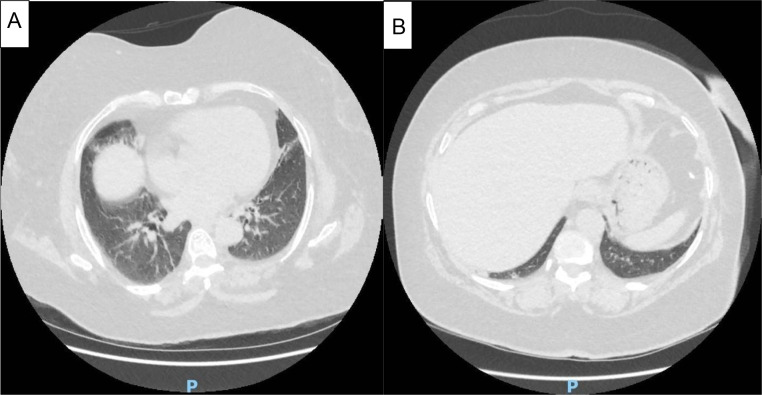
Rheumatoid arthritis is a chronic inflammatory rheumatic disease that can cause extra-articular manifestations, in particular pleuropulmonary involvement, which is the second leading cause of death in this connective tissue disease. We report a rare pulmonary manifestation of RA. This was a 58-year-old patient being followed for RA on leflunomide with 3 excavated pulmonary nodules measuring 13 mm, 10 mm and 6 mm, for which the diagnostic hypotheses of infection, pulmonary metastases and rheumatoid nodules had been put forward. The diagnosis of excavated pulmonary nodules in rheumatoid arthritis was made after aspiration of one of the nodules, which ruled out infectious and cancerous causes. These nodules regressed 6 months after leflunomide was stopped.
Keywords: Leflunomide; Pulmonary nodules; Rheumatoid arthritis.
© 2024 The Authors. Published by Elsevier Inc. on behalf of University of Washington.
Figures


References
-
- Antin-Ozerkis D, Evans J, Rubinowitz A, Homer RJ, Matthay RA. Pulmonary manifestations of rheumatoid arthritis. Clin Chest Mehed. 2010;31:451–478. - PubMed
-
- Juge PA, Dieudé P. Pneumopathies interstitielles diffuses au cours de la polyarthrite rhumatoïde. Revue du rhumatisme monographies. 2017;84:347–351.
-
- Patel R, Naik S, Amchentsev A, Saleh A. A rare cause of multiple cavitary nodules. Chest. 2009;136:306–309. - PubMed
Publication types
LinkOut - more resources
Full Text Sources
