

Small intestinal metastasis in a lung adenocarcinoma patient with concurrent EML4-ALK V3 and TP53 mutations after distinct responses to tyrosine kinase inhibitors: A case report
- PMID: 39430483
- PMCID: PMC11489313
- DOI: 10.1016/j.heliyon.2024.e38839
Small intestinal metastasis in a lung adenocarcinoma patient with concurrent EML4-ALK V3 and TP53 mutations after distinct responses to tyrosine kinase inhibitors: A case report
Abstract
Background: Although anaplastic lymphoma kinase tyrosine kinase inhibitors (ALK-TKIs) have improved the survival rates of lung cancer patients with ALK fusion mutations, their effectiveness varies significantly across different subtypes. We report a case of small intestine metastasis in a lung adenocarcinoma patient with co-occurring echinoderm microtubule-associated protein-like 4 (EML4)-ALK fusion variant 3 (V3) and tumor protein 53 (TP53) mutations after distinct responses to ALK-TKIs.
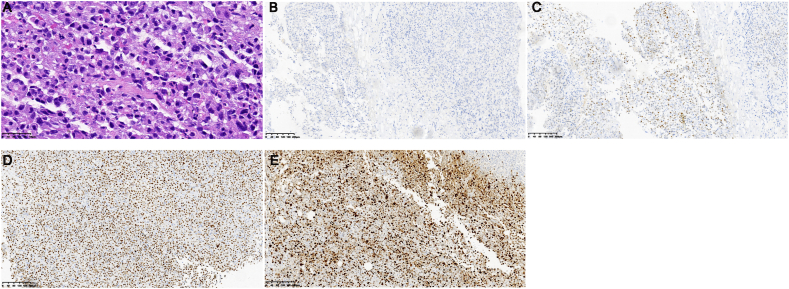
Case presentation: A 45-year-old woman was diagnosed with stage IV lung adenocarcinoma with brain metastasis. Next-generation sequencing revealed EML4-ALK V3 and TP53 co-mutations. After the initial treatment with ensartinib, the patient experienced intracranial disease progression. Radiation therapy (RT) was then administered. Despite good response to RT for the intracranial disease, the primary tumor enlarged. Thus, the patient was treated with oral ensartinib concurrent with chemotherapy, with a partial response in both the primary tumor and intracranial metastases. However, after three cycles of treatment, the patient discontinued chemotherapy because of acute kidney injury. Subsequent thoracic RT resulted in a partial response of the primary tumor; however, new brain and bone metastases were detected, prompting a switch to lorlatinib. The patient developed symptoms of intestinal obstruction 14 months after the initial diagnosis. Surgical intervention revealed a poorly differentiated metastatic lung adenocarcinoma of the upper jejunum. Genetic testing confirmed EML4-ALK V3 and TP53 co-mutations and high expression of programmed cell death-ligand 1. Despite pembrolizumab treatment, the patient's condition deteriorated, and she passed away.
Conclusion: We reported a rare case of small intestinal metastasis in a lung adenocarcinoma patient with concurrent EML4-ALK V3/TP53 mutations after distinct responses to ALK-TKIs in different lesions. Our findings revealed heterogeneity in ALK mutations and responses to ALK-TKIs, necessitating the close monitoring of genetic subtypes and associated mutations for tailored treatment strategies. Maintaining a heightened awareness of potential intestinal metastasis and vigilance in monitoring intestinal symptoms and abdominal metastases are pivotal for managing advanced lung adenocarcinoma.
Keywords: Anaplastic lymphoma kinase; Lorlatinib; Lung adenocarcinoma; Metastasis; Small intestine.
© 2024 The Authors.
Conflict of interest statement
The authors declare that they have no known competing financial interests or personal relationships that could have appeared to influence the work reported in this paper.
Figures


Similar articles
-
Ensartinib for EML4-ALK-positive lung adenocarcinoma with comorbid mutations in TP53, EGFR, and ERBB2: a case report.Front Oncol. 2025 Feb 14;15:1520287. doi: 10.3389/fonc.2025.1520287. eCollection 2025. Front Oncol. 2025. PMID: 40052122 Free PMC article.
-
ALK fusion small cell transformation of lung adenocarcinoma: A case report and literature review.Zhong Nan Da Xue Xue Bao Yi Xue Ban. 2024 Apr 28;49(4):628-636. doi: 10.11817/j.issn.1672-7347.2024.230506. Zhong Nan Da Xue Xue Bao Yi Xue Ban. 2024. PMID: 39019792 Free PMC article. Review. Chinese, English.
-
A case report of exceptional clinical response to chemoradiotherapy and tyrosine kinase inhibitors in a patient with EML4-ALK fusion variant 1 non-small cell lung cancer.Transl Lung Cancer Res. 2020 Dec;9(6):2500-2507. doi: 10.21037/tlcr-20-1212. Transl Lung Cancer Res. 2020. PMID: 33489810 Free PMC article.
-
Identification of a highly lethal V3+ TP53+ subset in ALK+ lung adenocarcinoma.Int J Cancer. 2019 Jan 1;144(1):190-199. doi: 10.1002/ijc.31893. Epub 2018 Oct 30. Int J Cancer. 2019. PMID: 30255938
-
Going beneath the tip of the iceberg. Identifying and understanding EML4-ALK variants and TP53 mutations to optimize treatment of ALK fusion positive (ALK+) NSCLC.Lung Cancer. 2021 Aug;158:126-136. doi: 10.1016/j.lungcan.2021.06.012. Epub 2021 Jun 12. Lung Cancer. 2021. PMID: 34175504 Review.
Cited by
-
Case Report: Intestinal metastasis from ALK-rearranged pulmonary pleomorphic carcinomas mimicking inflammatory myofibroblastic tumors.Front Oncol. 2025 Mar 28;15:1496752. doi: 10.3389/fonc.2025.1496752. eCollection 2025. Front Oncol. 2025. PMID: 40224190 Free PMC article.
References
-
- Bray F., Laversanne M., Sung H., Ferlay J., Siegel R.L., Soerjomataram I., et al. Global cancer statistics 2022: GLOBOCAN estimates of incidence and mortality worldwide for 36 cancers in 185 countries. Ca - Cancer J. Clin. 2024;74(3):229–263. - PubMed
-
- Thai A.A., Solomon B.J., Sequist L.V., Gainor J.F., Heist R.S. Lung cancer. Lancet. 2021;398(10299):535–554. - PubMed
-
- Jevremovic V. Is gastrointestinal metastasis of primary lung malignancy as rare as reported in the literature? A comparison between clinical cases and post-mortem studies. Oncology & Hematology Review (US) 2016;12:51.
-
- Stenbygaard L.E., Sørensen J.B. Small bowel metastases in non-small cell lung cancer. Lung Cancer. 1999;26(2):95–101. - PubMed
Publication types
LinkOut - more resources
Full Text Sources
Research Materials
Miscellaneous
