

Case Reports
doi: 10.1016/j.jdcr.2024.07.011.
eCollection 2024 Nov.
Relapsing toxic epidermal necrolysis following COVID-19
Affiliations
- PMID: 39430626
- PMCID: PMC11488411
- DOI: 10.1016/j.jdcr.2024.07.011
Item in Clipboard
Case Reports
Relapsing toxic epidermal necrolysis following COVID-19
JAAD Case Rep.
.
No abstract available
Keywords: COVID-19; SJS; Stevens-Johnson syndrome; TEN; relapsing; toxic epidermal necrolysis.
Conflict of interest statement
Dr Min is on the advisory boards of Horizon, McGraw Hill, and BMS and is an investigator for Amgen, BI, MBS, and Priovant. Dr Rojek has served on the advisory board of Boehringer Ingelheim. The other authors have no conflicts of interest to declare.
Figures
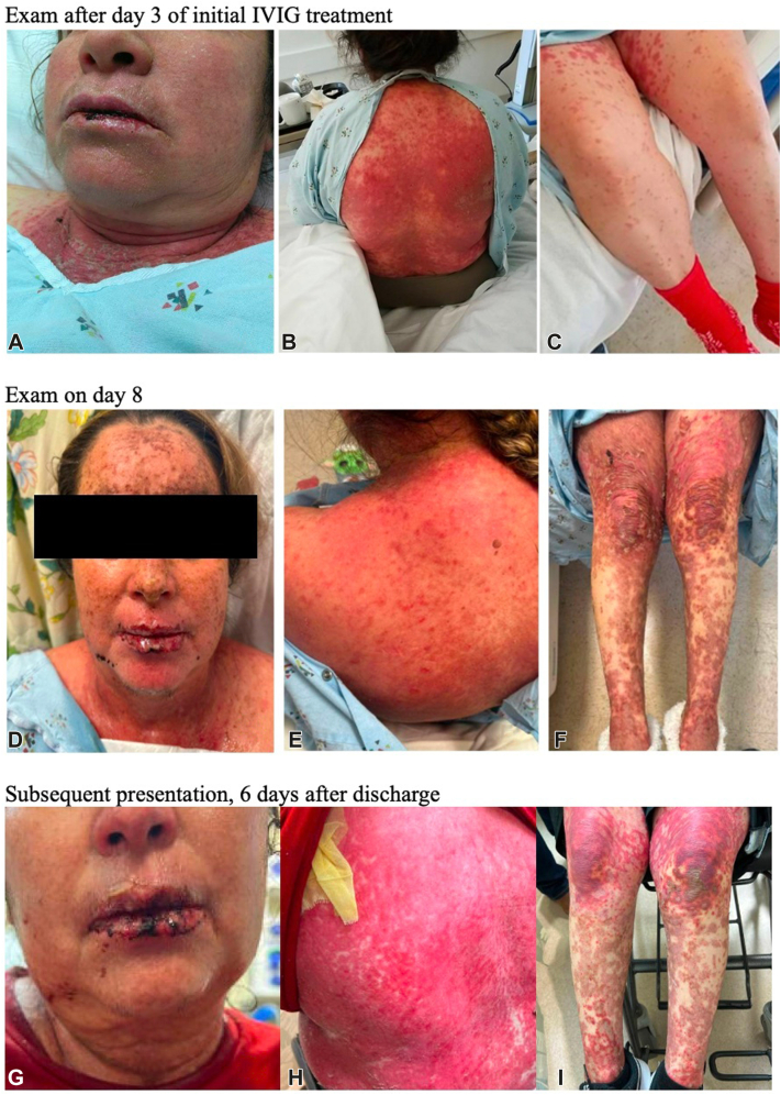
Examination after day 3 of initial IVIG treatment. Diffuse morbilliform eruption with bullae formation overlying dusky skin necrosis involved the (A) mucosal lips, neck, chest, (B) back, and (C) extremities. Examination on day 8. Dusky and necrosed papules coalescing into plaques with overlying flaccid and hemorrhagic bullae are seen on (D) the face and mucosal lips, (E) trunk including her back, and (F) extremities. Subsequent presentation, 6 days after discharge from initial presentation. G, Hemorrhagic crust on the lips. H, Diffuse erythroderma and erosions on the back with skin sloughing. I, Dusky papules coalescing into plaques with overlying flaccid and hemorrhagic bullae on the lower extremities.
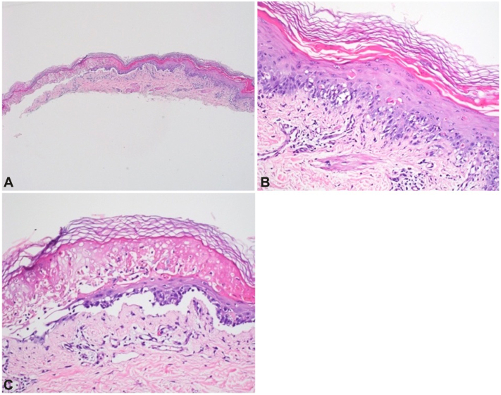
Shave biopsy revealed (A, C) a subepidermal split with areas of full-thickness epidermal necrosis and (B, C) interface dermatitis with scattered necrotic keratinocytes. (A-C, Hematoxylin-eosin stain; original magnifications: A, ×4; B and C, ×40.)
References
-
- Malviya M., Barua S., Shenoy M.A., Ullah A.M. Paxlovid-associated Stevens-Johnson syndrome. Chest J. 2023;164(4):A2062–A2063. doi: 10.1016/j.chest.2023.07.1410. - DOI
Publication types
LinkOut - more resources
Full Text Sources