. 2024 Oct 4:15:1470620.
doi: 10.3389/fimmu.2024.1470620. eCollection 2024.

Dynamic roles of neutrophil extracellular traps in cancer cell adhesion and activation of Notch 1-mediated epithelial-to-mesenchymal transition in EGFR-driven lung cancer cells

Affiliations

Dynamic roles of neutrophil extracellular traps in cancer cell adhesion and activation of Notch 1-mediated epithelial-to-mesenchymal transition in EGFR-driven lung cancer cells

Jelena Dimitrov et al. Front Immunol. .

Abstract

Introduction: Neutrophil extracellular traps (NETs) are complex structures released by activated neutrophils that may modulate different steps of the metastatic cascade. The aim of our study was to investigate how NETs can modulate the adhesion properties of cancer cells and whether cell exposure to NETs can activate the epithelial-to-mesenchymal transition (EMT) program thus enhancing the migratory and invasive properties of tumor cells.

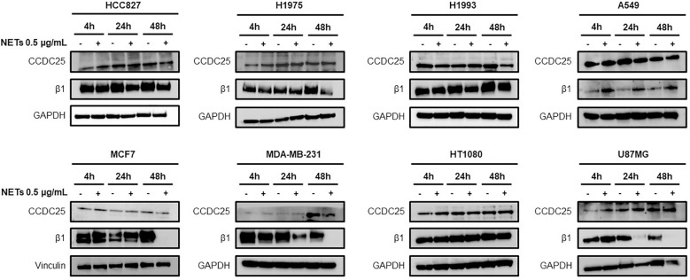
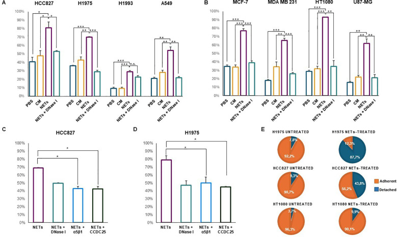
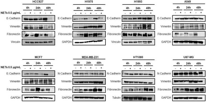
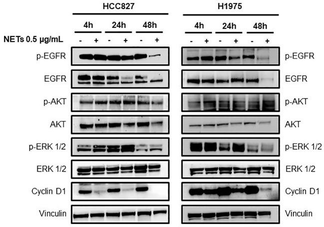
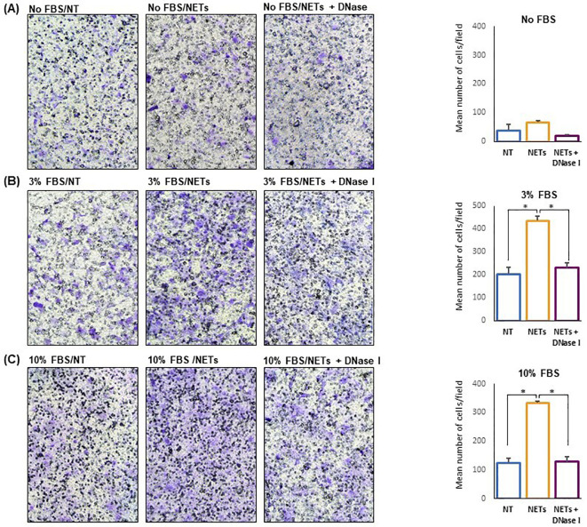
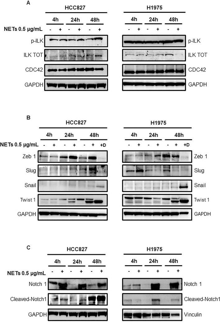
Materials and methods: Different cancer cell lines were subjected to a solid-phase adhesion assay using NET-coated plates with or without the addition of antibodies against α5β1 or CCDC25 receptor. After 1-4 h of incubation, adherent cells were expressed as the percentage of total cell number. To test EMT occurrence, cells were treated with NETs for up to 48 h and then the levels of E-cadherin, vimentin, Snail, Slug, Zeb 1 and Twist 1 along with levels of Notch 1 and cleaved Notch 1 were determined by western blotting. Untreated and NET-treated cells were subjected to migration assays using 24-multiwell plates with transwell and FBS as chemoattractant.

Results: Cancer cell adhesion to NET-coated plates varied between 30% and 92.7% and was significantly higher than that obtained in uncoated plates. The addition of antibodies against α5β1 or CCDC25 caused a strong reduction of cell adhesion to NETs. The prolonged exposure of EGFR-driven cancer cell lines to NETs caused the activation of the EMT program through the upregulation and cleavage of Notch 1 and was confirmed by the enhanced expression of EMT markers. The consequent loss of the epithelial phenotype induced a strong reduction of the expression of the oncogene driver. Cell migration was significantly enhanced in NET-treated cells as compared to untreated cells.

Discussion: Our findings reveal the dynamic role of NETs that may provide a DNA and fibronectin rich environment for binding of many cancer cells at distant sites where the prolonged exposure to NETs triggers the EMT through the activation of Notch 1 signaling pathway with the subsequent enhancement of migratory and invasive properties of cancer cells. Furthermore, our findings provide an example of how an immune/inflammatory microenvironment may directly modulate the sensitivity of cancer cells to oncogene targeted agents.

Keywords: Notch 1; adhesion; epithelial mesenchymal transition; metastasis; migration; neutrophil extracellular traps; tumor microenvironment.


Conflict of interest statement

The authors declare that the research was conducted in the absence of any commercial or financial relationships that could be construed as a potential conflict of interest.

Figures

Figure 1
Figure 1
Levels of CCDC25 and β1 chain expression in different cancer cell lines. Cells were exposed or not to 0.5 µg/mL NETs for 4 h, 24 h and 48 h and then subjected to western blot analysis. GAPDH or vinculin were used as equal loading.
Figure 2
Figure 2
Cell adhesion of different cancer cell lines in the presence or absence of NETs. Cells were seeded and allowed to adhere to NET-coated multi-well plates for 1, 2 or 4 h depending on the cell line (A–D). Negative controls were plates coated with PBS or conditioned medium of dHL-60 and NET-coated plates pre-incubated with DNase I. Results are expressed as percentage of total cell number in each well. In parallel experiments, adherent cells were exposed to 0.5 µg/mL NETs for 48 h followed by counting of adherent and detached viable cells (E). (A) Cell adhesion (mean ± SE) obtained in HCC827 (2 h), H1975 (2 h), H1993 (2 h) and A549 (1 h) lung cancer cell lines. (B) Cell adhesion (mean ± SE) obtained in MCF7 (2 h) and MDA MB231 (2 h) breast cancer cells, HT1080 fibrosarcoma (1 h) and U87-MG glioblastoma (4 h) cell lines. (C) Cell adhesion (mean ± SE) to NET-coated plates obtained in HCC827 cells pre-incubated with blocking antibodies against α5β1 integrin and CCDC25 receptor. (D) Cell adhesion (mean ± SE) to NET-coated plates obtained in H1975 cells pre-incubated with blocking antibodies against α5β1 integrin and CCDC25 receptor. (E) Percentage of adherent and detached viable cells exposed or not to NETs in the medium for 48 h. Statistical significance: *p<0.05; **p< 0.01; ***p< 0.001; ns, not significant.
Figure 3
Figure 3
Expression of EMT markers in response to NET treatment. Levels of E-cadherin, vimentin, N-cadherin and fibronectin in different cancer cell lines exposed or not to 0.5 µg/mL NETs for 4 h, 24 h and 48 h. GADPH, vinculin and tubulin were used as equal loading.
Figure 4
Figure 4
Loss of epithelial phenotype in EGFR-driven lung cancer cells exposed to NETs. HCC827 and H1975 cells were incubated with 0.5 µg/mL NETs for 4 h, 24 h and 48 h. Levels of p-EGFR, EGFR, p-AKT, AKT, p-ERK 1/2, ERK 1/2 and cyclin D1 were determined. Vinculin were used as equal loading.
Figure 5
Figure 5
NET-dependent enhancement of cell migration. Untreated or NET-treated H1975 cells were seeded in the upper compartment of transwell and allowed to migrate for 16 h. No chemoattractant, 3% FBS or 10%FBS was added to the medium in the lower compartment. On the left, representative images showing migrated cells (100X Magnification) obtained with no FBS (A), 3% FBS (B) and 10% FBS (C) in the lower compartment. On the right, the corresponding graph showing the mean number/field (± SE) of migrated cells obtained in each condition. Cell migration was 1.7 (A), 2.1 (B), and 2.7 (C) folds higher in NET-treated cells as compared to untreated cells when medium without serum, or supplemented with 3% FBS and 10% FBS, respectively, were used as chemoattractant. Statistical significance: *p<0.05.
Figure 6
Figure 6
Levels of signaling mediators in EGFR-driven HCC827 and H1975 cells exposed to NETs. (A) Levels of p-ILK, ILK and CDC42 in cells exposed or not to 0.5 µg/mL NETs for 4 h, 24 h and 48 h. (B) Levels of transcription factors in adherent cells exposed or not to treatment with NETs for 4 h, 24 h and 48 h (lanes 2,4,6) and in viable cells that were detached after 48 h treatment with NETs (lane 7, +D). (C) Levels of total Notch 1 and cleaved Notch 1 in HCC827 and H1975 cells exposed or not to NETs for 4 h, 24 h and 48 h. GAPDH and vinculin were used as equal loading.

References

    1. Brinkmann V, Reichard U, Goosmann C, Fauler B, Uhlemann Y, Weiss DS, et al. . Neutrophil extracellular traps kill bacteria. Science. (2004) 303:1532–5. doi: 10.1126/science.1092385 - DOI - PubMed
    1. Papayannopoulos V. Neutrophil extracellular traps in immunity and disease. Nat Rev Immunol. (2018) 18:134–47. doi: 10.1038/nri.2017.105 - DOI - PubMed
    1. Fuchs TA, Abed U, Goosmann C, Hurwitz R, Schulze I, Wahn V, et al. . Novel cell death program leads to neutrophil extracellular traps. J Cell Biol. (2007) 176:231–41. doi: 10.1083/jcb.200606027 - DOI - PMC - PubMed
    1. Demers M, Krause DS, Schatzberg D, Martinod K, Voorhees JR, Fuchs TA, et al. . Cancers predispose neutrophils to release extracellular DNA traps that contribute to cancer-associated thrombosis. Proc Natl Acad Sci USA. (2012) 109:13076–81. doi: 10.1073/pnas.1200419109 - DOI - PMC - PubMed
    1. Masucci MT, Minopoli M, Del Vecchio S, Carriero MV. The emerging role of neutrophil extracellular traps (Nets) in tumor progression and metastasis. Front Immunol. (2020) 11:1749. doi: 10.3389/fimmu.2020.01749 - DOI - PMC - PubMed