. 2024 Oct 2:42:100879.
doi: 10.1016/j.bbih.2024.100879. eCollection 2024 Dec.

Early-life obesogenic environment integrates immunometabolic and epigenetic signatures governing neuroinflammation

Affiliations

Early-life obesogenic environment integrates immunometabolic and epigenetic signatures governing neuroinflammation

Perla Ontiveros-Ángel et al. Brain Behav Immun Health. .

Abstract

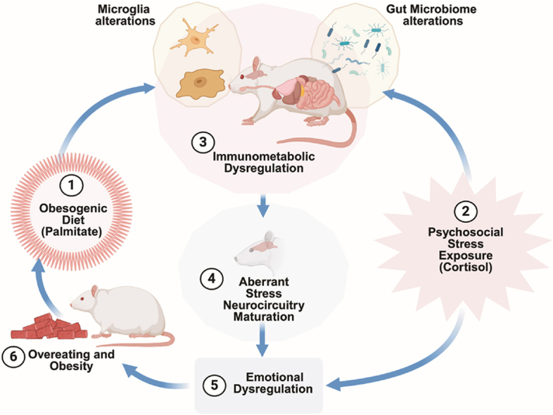
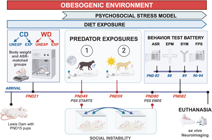
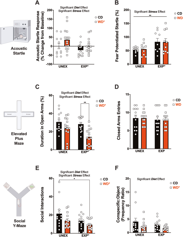
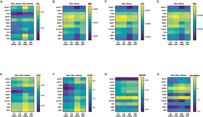
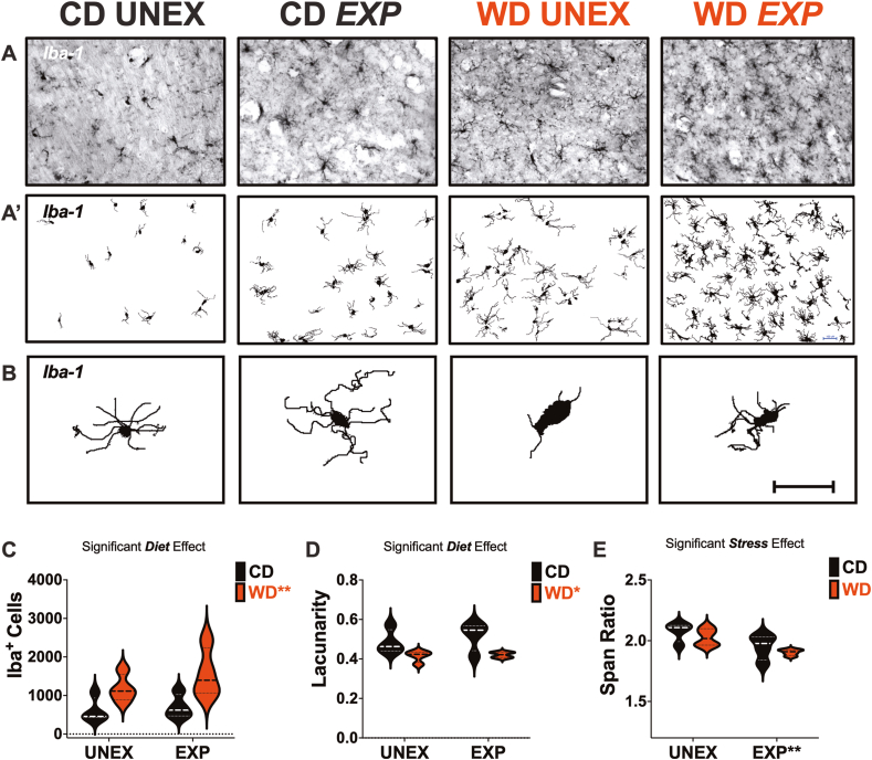
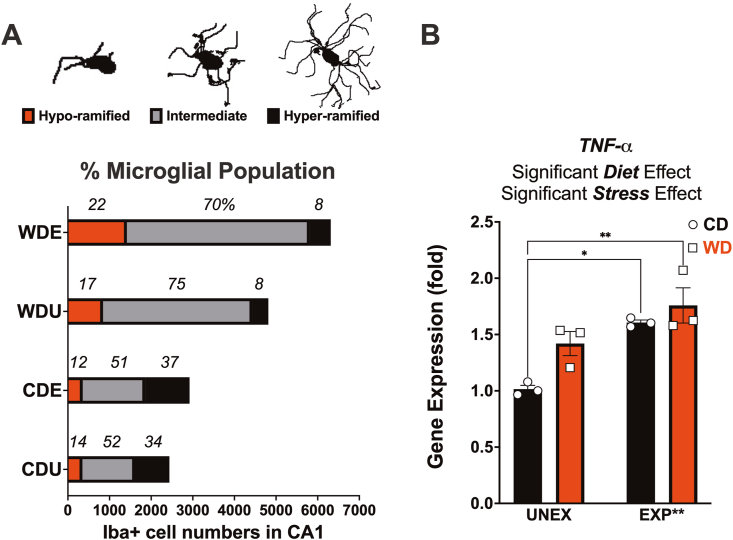
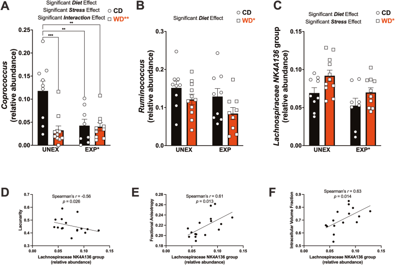
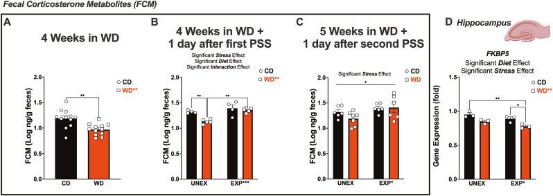
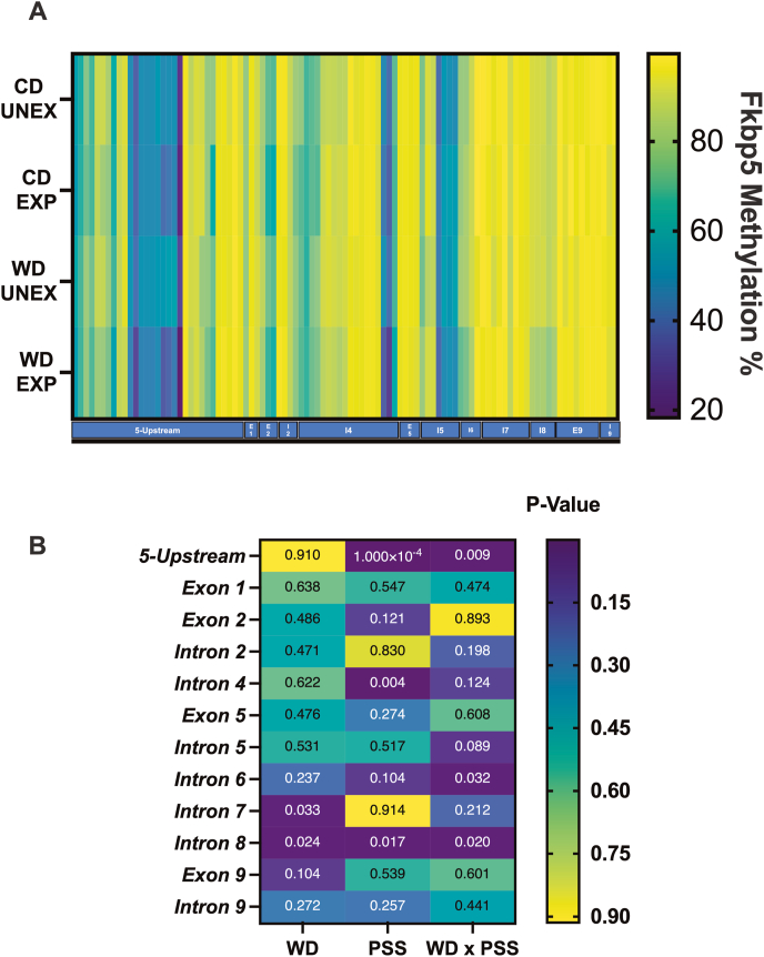
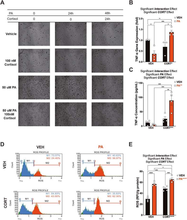
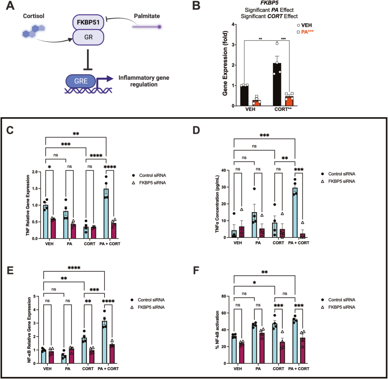
Childhood overweight/obesity is associated with stress-related psychopathology, yet the pathways connecting childhood obesity to stress susceptibility are poorly understood. We employed a systems biology approach with 62 adolescent Lewis rats fed a Western-like high-saturated fat diet (WD, 41% kcal from fat) or a control diet (CD, 13% kcal from fat). A subset of rats underwent a 31-day model of predator exposures and social instability (PSS). Effects were assessed using behavioral tests, DTI (diffusion tensor imaging), NODDI (neurite orientation dispersion and density imaging), 16S rRNA gene sequencing for gut microbiome profiling, hippocampal microglia analysis, and targeted gene methylation. Parallel experiments on human microglia cells (HMC3) examined how palmitic acid influences cortisol-related inflammatory responses. Rats exposed to WD and PSS exhibited deficits in sociability, increased fear/anxiety-like behaviors, food consumption, and body weight. WD/PSS altered hippocampal microstructure (subiculum, CA1, dentate gyrus), and microbiome analysis showed a reduced abundance of members of the phylum Firmicutes. WD/PSS synergistically promoted neuroinflammatory changes in hippocampal microglia, linked with microbiome shifts and altered Fkbp5 expression/methylation. In HMC3, palmitate disrupted cortisol responses, affecting morphology, phagocytic markers, and cytokine release, partially mediated by FKBP5. This study identifies gene-environment interactions that influence microglia biology and may contribute to the connection between childhood obesity and stress-related psychopathology later in life.

Keywords: Adolescence; Anxiety; FKBP5; Microbiome; Microglia; NODDI; Neuroinflammation; Obesity.


Conflict of interest statement

The authors declare no conflicts of interest related to this work.

Figures

Image 1
Graphical abstract
Fig. 1
Fig. 1
Experimental timeline and procedures. Adolescent Lewis rats (PND21) were grouped based on body weight and acoustic startle reactivity and assigned to either a Western-like high-saturated fat diet or a purified control diet with matched ingredients. These diets were consumed for four weeks, after which the rats were divided into four groups through random selection at PND49: (1) control diet, unexposed (CDU); (2) control diet, exposed (CDE); (3) Western diet, unexposed (WDU); (4) Western diet, exposed (WDE); each group comprising n = 14 rats. Exposed groups underwent a well-established 31-day psychosocial stress (PSS) model involving predator encounters and social instability. At PND82, the effects of WD and PSS were evaluated using a comprehensive battery of standard behavioral tests and various measures of stress and inflammation markers. Subsequently, all rats were euthanized, and plasma and brain tissue samples were collected. Abbreviations: ASR, Acoustic startle reflex, EPM, Elevated plus maze; SYM, Social Y-Maze; FPS, Fear-potentiated startle.
Fig. 2
Fig. 2
Adolescent access to an obesogenic diet (WD) and psychosocial stress (PSS) influence startle reactivity, anxiety-like behavior, and indices of sociability. (A) PSS (p = 0.035) and WD (p = 0.025) had opposing influences on the change in ASR responses from baseline. (B) PSS increased the fear-potentiated startle (FPS) (p = 0.0097). (C) WD consumption (p = 0.0002) and PSS exposure (p = 0.033) significantly reduce time spent in the open arms of the elevated plus maze. Post hoc revealed that WD rats exposed to PSS significantly decreased the time spent in the OA compared to the CD EXP rats (p = 0.0024). (D) The number of closed arms entries (a proxy of motor activity in the maze) was not affected by the experimental conditions (p > 0.0050). (E) WD (p = 0.0273) and PSS (p = 0.0256) reduced the number of social interactions in the social Y-Maze. Post hoc revealed that the number of social interactions was robustly decreased by the combined effects of the WD and PSS (p = 0.0134 relative to control). (F) The WD reduced the frequency ratio of interactions between the conspecific and the object (p = 0.047). The sample size for all behavioral tests was 14 rats per group (before outlier testing).
Fig. 3
Fig. 3
Exposure to an obesogenic environment during adolescence impacts the structural integrity of distinctive hippocampal subfields, subregions, and adjacent areas. Heatmaps of hippocampal structural changes based on each diffusion-MRI parameter value. (A) Fractional anisotropy (FA) was significantly modified by the WD (p < 0.0001), PSS (p = 0.0022), and the interactions between WD and PSS (p = 0.022). (BD) Medial (MD), axial (AD), and radial diffusivity (RD) were also significantly altered by the WD (MD, p = 0.030; AD, p = 0.036; RD, p = 0.028) and PSS (MD, p = 0.030; AD, p = 0.033; RD, p = 0.030). (EH) Orientation dispersion index (ODI), intracellular volume fraction (ICVF), and Jacobian lob matrix data derived from DTI/NODDI analysis revealed that the WD (ODI, p < 0.0001; ICVF, p < 0.0001; Jacobian, p = 0.0023), and WD x PSS (p = 0.0131) (p = 0.0057) (p = 0.0270) significantly contributed to changes in hippocampal tissue integrity. (G) The isotropic volume fraction remained unaffected by the experimental conditions. This indicates that changes observed in other DTI metrics are not due to alterations in the overall isotropic volume but may instead reflect changes in tissue microstructure or organization. Asterisks indicate region-specific post hoc effects that significantly contribute to the main and interaction effects observed in the two-way ANOVA (FA: dCA1, vCA1; MD: dCA1, dDG, vSb; AD: dCA1, dDG, dSb, vSb; RD: dCA1, dDG, vSb; ODI: dDG, vDG, vSb; ICVF: vCA1, vDG, dSb, vSb, Jabobian: vCA1, dSb). Sample numbers: CD UNEXP, n = 6; CD EXP, n = 7; WD UNEXP, n = 8, WD EXP; n = 7. Abbreviations: d, dorsal; v, ventral; CA, Cornu Ammonis, DG, dentate gyrus, EcrhC, ectorhinal cortex; EC, entorhinal cortex; Sb, subiculum; CD, control diet; WD, Western-like diet; UNEX, unexposed; EXP, PSS exposed.
Fig. 4
Fig. 4
An obesogenic environment promotes microglial proliferation and proinflammatory morphology in the rat hippocampus. (A) Representative images of full-focused composite 10 × brightfield images from hippocampal CA1 were acquired and stained for Iba-1. (A′) Corresponding binarized images were processed for quantitative morphometric analysis. (B) Representative microglia morphologies in each study group. (C) The WD significantly increased the total Iba1+ cells (p = 0.0056). (D) Microglial lacunarity was significantly reduced by the WD (p = 0.0057). (E) Microglial span ratio was significantly reduced in PSS-exposed rats (p = 0.0064). Sample size = 4 rats/group. Scale bar for all images: 100 μm.
Fig. 5
Fig. 5
Obesogenic conditions in adolescence promote significant expansion of 'intermediate' microglia phenotype and TNF-α expression. Principal component analysis (PCA) and K-means clustering were used to generate three unique microglial phenotypes based on lacunarity. (A) Representative images of the three distinct morphological phenotypes: Hyper-ramified, Intermediate, and Hypo-ramified. The experimental conditions significantly altered the microglial cell distribution based on lacunarity (n, χ2, df, p; 6,49, 1982, 6, p < 0.0001). Hypo-ramified and intermediate phenotypes increased in response to the WD and PSS, while the amount of hyper-ramified cells was reduced. (B) Hippocampal TNF-α gene expression was shown to be significantly influenced by both the WD (p = 0.021) and PSS (p = 0.0014). Relative to UNEX controls, PSS exposure significantly increased hippocampal TNF-α gene expression in CD (p = 0.0110) and WD (p = 0.0028) fed rats. Sample size: histology: 4 rats/group, 16,487 Iba-1+ cells; RT-PCR: 3 rats/group.
Fig. 6
Fig. 6
An obesogenic diet and chronic psychosocial stress induce alterations in Firmicutes linked to changes in microglia morphology and brain microstructure. (A) Microbiome evaluation revealed that the WD (p = 0.0033), PSS (p = 0.021), and interaction of WD and PSS (p = 0.0052) significantly contributed to the relative abundance of Coprococcus in the gut. Multiple comparisons analysis revealed that, relative to controls, there was a significant reduction in Coprococcus abundance due to the WD (p = 0.0005), PSS (p = 0.0048), and the combined effect of WD and PSS (p = 0.0019). (B) The WD significantly reduced the abundance of Ruminococcus (p = 0.035). (C) The WD increased (p = 0.012), while PSS decreased (p = 0.015), the relative abundance of Lachnospiraceae NK4A136. Sample numbers: CD UNEXP, n = 9; CD EXP, n = 11; WD UNEXP, n = 8, WD EXP; n = 10. Spearman's rank correlation analysis showed significant associations between Lachnospiraceae NK4A136 abundance and lacunarity (D), fractional anisotropy (E), and intracellular volume fraction (F)—correlation sample size = 12 rats. Detailed information on microbiota analyses, correlations, false discoveries, and other related statistics can be found in the Supplementary Materials.
Fig. 7
Fig. 7
Exposure to an obesogenic diet and chronic psychosocial stress influence corticosterone levels and hippocampal Fkbp5 gene expression. (A) Fecal corticosterone metabolite (FCM) levels were significantly decreased by the WD (p = 0.0018). (B) We observed significant effects of WD (p = 0.0052), PSS (p = 0.0008), and their interaction (p = 0.039) following the first day after predatory exposure. PSS significantly increased FCM levels in the WD rats (p = 0.0012). (C) FCM remained significantly elevated in PSS rats 24 h after the second predatory exposure (p = 0.0250). (D) Hippocampal Fkbp5 mRNA levels were significantly decreased by the WD (p = 0.0016) and PSS (p = 0.020). RT-PCR: 3 rats/group.
Fig. 8
Fig. 8
Heatmap and analysis of rat Fkbp5 methylation signatures per gene region. (A) Global percentages of Fkbp5 methylation per locus (B) Breakdown of significance per region. Methylation in the 5′-upstream region was significantly modified by PSS (p = 0.0001) and the interaction of WD x PPS (p = 0.009). Intron 4 was significantly modified by PSS (p = 0.003). Intron 6 was modified by WD x PSS (p = 0.032). Intron 7 was modified by the diet (p = 0.032); Intron 8 was modified by WD (p = 0.024), PSS (p = 0.017), and WD x PSS (0.024). The summary of statistical values of multiple comparisons in FKBP5 by regions can be found in Supplementary Table 16A–C and 17.
Fig. 9
Fig. 9
Palmitic acid sensitizes human microglial cells to abnormal responses to cortisol. (A) Representative brightfield images of human microglial cells (HCM3 cells) pretreated with either vehicle (VEH) or 50 μM palmitic acid (PA), followed by treatment with VEH or 100 nM hydrocortisone (CORT) 24 h later. Cells and supernatant were collected 24 h after CORT/VEH treatment. (B) We found an interaction effect on TNF-α mRNA levels (p = 0.0002), indicating that the relationship between PA and TNF-α mRNA expression depends on the presence of CORT. Incubation of PA followed by CORT promoted a dramatic increase in TNF-α mRNA expression relative to control conditions (p = 0.0003). (C) TNF-α protein levels were also significantly influenced by PA (p = 0.0096), CORT (p = 0.0002), and the interaction of PA x CORT (p = 0.0021), partly supporting the mRNA expression results. (D) Reactive Oxygen Species (ROS -intracellular superoxide levels) were detected by dihydroethidium (DHE) measurements, showing increased ROS-positive cells after PA (33% increase) and CORT (19% increase) treatment. PA priming resulted in a 38% increase in ROS + cells after CORT treatment (relative to control conditions). M1 = ROS negative cells; M2 = ROS positive cells. PA increased (E) Quantification of ROS levels normalized by grams of protein demonstrated a significant ROS increase in PA (p < 0.0001) and CORT (p < 0.0001) treated cells. We also observed a significant interaction effect (p = 0.0459). RFU: relative fluorescence unit. ∗p < 0.05, ∗∗p < 0.01 and ∗∗∗p < 0.001 ∗∗∗∗p < 0.0001. Sample size = four independent experiments. Error bars represent standard errors.
Fig. 10
Fig. 10
Human FKBP5 regulates microglial TNF-α levels under obesogenic factors. (A) Schematic model illustrating the primary hypothesis that PA disrupts pro-inflammatory gene expression mediated by the glucocorticoid receptor (GR) through FKBP5/FKBP51 regulation. (B) In support of this notion, PA significantly reduced FKBP5 mRNA levels (p < 0.0001)—sample size = four independent experiments. FKBP5 siRNA delivery attenuated the impact of PA + CORT on TNF-α upregulation (p < 0.0001) (C) and released protein levels of TNF-α (p = 0.0001) (D). CORT increased NF-κB gene expression (E) and protein activation (p < 0.05) (F). This effect was reduced in cells treated with FKBP5 siRNA (p < 0.001) (E-F)—sample size = four independent experiments.

References

    1. Abawi O., Welling M.S., Eynde E., Rossum E.F.C., Halberstadt J., Akker E.L.T., et al. COVID‐19 related anxiety in children and adolescents with severe obesity: a mixed‐methods study. Clin Obes. 2020;10 - PMC - PubMed
    1. Adamec R.E., Shallow T. Lasting effects on rodent anxiety of a single exposure to a cat. Physiol. Behav. 1993;54:101–109. - PubMed
    1. Adamec R., Strasser K., Blundell J., Burton P., McKay D.W. Protein synthesis and the mechanisms of lasting change in anxiety induced by severe stress. Behav. Brain Res. 2006;167:270–286. - PubMed
    1. Asch R.H., Holmes S.E., Jastreboff A.M., Potenza M.N., Baldassarri S.R., Carson R.E., et al. Lower synaptic density is associated with psychiatric and cognitive alterations in obesity. Neuropsychopharmacology. 2021:1–10. - PMC - PubMed
    1. Bakoyiannis I., Ducourneau E.G., N’diaye M., Fermigier A., Ducroix-Crepy C., Bosch-Bouju C., et al. Obesogenic diet induces circuit-specific memory deficits in mice. Elife. 2024;13 - PMC - PubMed