. 2024 Oct 3:12:1475918.
doi: 10.3389/fbioe.2024.1475918. eCollection 2024.

Effective removal of host cell-derived nucleic acids bound to hepatitis B core antigen virus-like particles by heparin chromatography

Affiliations

Effective removal of host cell-derived nucleic acids bound to hepatitis B core antigen virus-like particles by heparin chromatography

Angela Valentic et al. Front Bioeng Biotechnol. .

Abstract

Virus-like particles (VLPs) show considerable potential for a wide array of therapeutic applications, spanning from vaccines targeting infectious diseases to applications in cancer immunotherapy and drug delivery. In the context of hepatitis B core antigen (HBcAg) VLPs, a promising candidate for gene delivery approaches, the naturally occurring nucleic acid (NA) binding region is commonly utilized for effective binding of various types of therapeutic nucleic acids (NAther). During formation of the HBcAg VLPs, host cell-derived nucleic acids (NAhc) might be associated to the NA binding region, and are thus encapsulated into the VLPs. Following a VLP harvest, the NAhc need to be removed effectively before loading the VLP with NAther. Various techniques reported in literature for this NAhc removal, including enzymatic treatments, alkaline treatment, and lithium chloride precipitation, lack quantitative evidence of sufficient NAhc removal accompanied by a subsequent high VLP protein recovery. In this study, we present a novel heparin chromatography-based process for effective NAhc removal from HBcAg VLPs. Six HBcAg VLP constructs with varying lengths of the NA binding region and diverse NAhc loadings were subjected to evaluation. Process performance was thoroughly examined through NAhc removal and VLP protein recovery analyses. Hereby, reversed phase chromatography combined with UV/Vis spectroscopy, as well as silica spin column-based chromatography coupled with dye-based fluorescence assay were employed. Additionally, alternative process variants, comprising sulfate chromatography and additional nuclease treatments, were investigated. Comparative analyses were conducted with LiCl precipitation and alkaline treatment procedures to ascertain the efficacy of the newly developed chromatography-based methods. Results revealed the superior performance of the heparin chromatography procedure in achieving high NAhc removal and concurrent VLP protein recovery. Furthermore, nuanced relationships between NA binding region length and NAhc removal efficiency were elucidated. Hereby, the construct Cp157 surpassed the other constructs in the heparin process by demonstrating high NAhc removal and VLP protein recovery. Among the other process variants minimal performance variations were observed for the selected constructs Cp157 and Cp183. However, the heparin chromatography-based process consistently outperformed other methods, underscoring its superiority in NAhc removal and VLP protein recovery.

Keywords: HBcAg; heparin chromatography; host cell-derived nucleic acids; process development; virus-like particles.


Conflict of interest statement

The authors declare that the research was conducted in the absence of any commercial or financial relationships that could be construed as a potential conflict of interest.

Figures

FIGURE 1
FIGURE 1
(A) Common HBcAg VLP purification process (Hillebrandt et al., 2021) with depletion of NAhc and loading with NAther, process steps required for the application as a nucleic acid delivery vector, highlighted in blue. The depletion of NAhc investigated in this study is highlighted in grey. (B) HBcAg VLP with NA binding region and NAhc encapsulated within the VLP protein capsid. (C) HBcAg VLP with NA binding region and NAther encapsulated within the VLP protein capsid after depletion of NAhc and loading with NAther. HBcAg: hepatitis B core antigen, NA: nucleic acids, NAhc: host cell-derived nucleic acids, NAther: therapeutic nucleic acids, VLP: virus-like particle.
FIGURE 2
FIGURE 2
Overview of processes investigated in this work to remove host cell-derived nucleic acids bound to HBcAg VLPs. Heparin and sulfate chromatography were performed with and without prior nuclease treatment. LiCl precipitation and alkaline treatment were adapted from reported techniques in literature (Porterfield et al., 2010; Petrovskis et al., 2021). The alkaline treatment procedure was performed with and without the reported preliminary dialysis in 7 M Urea and NaCO3 solutions. All outlined removal techniques were performed with Cp157 and Cp183 HBcAg VLP constructs (Valentic et al., 2022), while the grey shaded process path was additionally performed for Cp149, Cp154, Cp164, and Cp167 (*). Initial, intermediate and final samples, which are labelled at the process steps, respectively, were analysed by SDS-PAGE, NAGE, RP chromatography coupled with UV/Vis analysis, and silica-SC based chromatography followed by dye-based fluorescence assay (Valentic et al., 2024). HBcAg: hepatitis B core antigen, NAGE: native agarose gel electrophoresis, NT: nuclease treatment, RP: reversed phase, VLP: virus-like particle.
FIGURE 3
FIGURE 3
Protein and nucleic acid recoveries for six HBcAg VLP constructs after removal of host cell-derived nucleic acids by disassembly and heparin chromatography (highlighted process path in Figure 1). Nucleic acid recoveries were analysed by silica-SC based chromatography followed by dye-based fluorescence assay and RP chromatography coupled with UV/Vis analysis. VLP protein recoveries were determined by RP chromatography coupled with UV/Vis analysis. HBcAg: hepatitis B core antigen, NAGE: native agarose gel electrophoresis, RP: reversed phase, SC: spin column, VLP: virus-like particle.
FIGURE 4
FIGURE 4
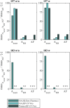
VLP protein and nucleic acid recoveries for Cp157 and Cp183 after removal of host cell-derived nucleic acids by disassembly, with (w) or without (w/o) prior nuclease treatment and (A) heparin chromatography or (B) sulfate chromatography. Nucleic acid recoveries were analysed by silica-SC based chromatography followed by dye-based fluorescence assay and RP chromatography coupled with UV/Vis analysis. VLP protein recoveries were determined by RP chromatography coupled with UV/Vis analysis. RP: reversed phase, SC: spin column.
FIGURE 5
FIGURE 5
VLP protein and nucleic acid recoveries for Cp157 and Cp183 after removal of host cell-derived nucleic acids by disassembly and LiCl precipitation, centrifugation (intermediate sample) and size-exclusion chromatography. Nucleic acid recoveries were analysed by silica-SC based chromatography followed by dye-based fluorescence assay and RP chromatography coupled with UV/Vis analysis. VLP protein recoveries were determined by RP chromatography coupled with UV/Vis analysis. Nucleic acid quantification by RP-UV/Vis was not possible after centrifugation due to the presence of guanidine HCl in the samples, overlapping with the nucleic acid peaks in the flow-through of the RP-HPLC method, and nucleic acid and VLP protein concentrations of Cp183 in SEC fraction were too low for RP-UV/Vis analysis (*). RP: reversed phase, SC: spin column, SEC: size-exclusion chromatography.
FIGURE 6
FIGURE 6
VLP protein and nucleic acid recoveries for Cp157 and Cp183 after removal of host cell-derived nucleic acids by alkaline treatment (dialysis in pH 12 buffer) with (w) and without (w/o) preliminary dialysis in 7 M Urea and NaCO3 solutions, and dialysis in neutralisation buffer (intermediate sample), precipitation and redissolution (intermediate sample) and size-exclusion chromatography. Nucleic acid recoveries were analysed by silica-SC based chromatography followed by dye-based fluorescence assay and RP chromatography coupled with UV/Vis analysis. VLP protein recoveries were determined by RP chromatography coupled with UV/Vis analysis. Nucleic acid and VLP protein concentrations of Cp183 for samples after dialysis in restoration buffer and redissolution (both without and with prior preliminary dialysis), were too low for RP-UV/Vis analysis, and concentrations during SEC were too low to collect and analyse fractions (*). RP: reversed phase, SC: spin column.
FIGURE 7
FIGURE 7
(A) Ionic stabilization of the NA binding region of the Cp157 and Cp183 HBcAg VLP constructs after removal of NAhc by NaCl. (B) Degradation of NA parts bound to the NA binding region by nucleases impeded by steric hindrance. HBcAg: hepatitis B core antigen, NA: nucleic acids, NAhc: host cell-derived nucleic acids, VLP: virus-like particle.

References

    1. Baytas S. N., Linhardt R. J. (2020). Advances in the preparation and synthesis of heparin and related products. Drug Discov. Today 25, 2095–2109. 10.1016/j.drudis.2020.09.011 - DOI - PMC - PubMed
    1. Begic M., Pecenkovic S., Gajdosik M. S., Josic D., Müller E. (2021). Salt‐tolerant cation exchanger‐containing sulfate groups as a viable alternative for mixed-mode type and heparin-based affinity resins. Biotechnol. J. 16, e2100100. 10.1002/biot.202100100 - DOI - PubMed
    1. Birnbaum F., Nassal M. (1990). Hepatitis B virus nucleocapsid assembly: primary structure requirements in the core protein. J. Virol. 64, 3319–3330. 10.1128/jvi.64.7.3319-3330.1990 - DOI - PMC - PubMed
    1. Boisgérault F., Morón G., Leclerc C. (2002). Virus-like particles: a new family of delivery systems. Expert Rev. Vaccines 1, 101–109. 10.1586/14760584.1.1.101 - DOI - PubMed
    1. Broos K., Vanlandschoot P., Maras M., Robbens J., Leroux-Roels G., Guisez Y. (2007). Expression, purification and characterization of full-length RNA-free hepatitis B core particles. Protein Expr. Purif. 54, 30–37. 10.1016/j.pep.2007.02.006 - DOI - PubMed

LinkOut - more resources