. 2024 Jan-Dec:23:15330338241288751.
doi: 10.1177/15330338241288751.

Radiomic Machine Learning in Invasive Ductal Breast Cancer: Prediction of Ki-67 Expression Level Based on Radiomics of DCE-MRI

Affiliations

Radiomic Machine Learning in Invasive Ductal Breast Cancer: Prediction of Ki-67 Expression Level Based on Radiomics of DCE-MRI

Huan Yang et al. Technol Cancer Res Treat. 2024 Jan-Dec.

Abstract

Purpose: Our study aimed to investigate the potential of radiomics with DCE-MRI for predicting Ki-67 expression in invasive ductal breast cancer.

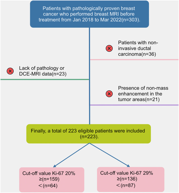
Method: We conducted a retrospective study including 223 patients diagnosed with invasive ductal breast cancer. Radiomics features were extracted from DCE-MRI using 3D-Slicer software. Two Ki-67 expression cutoff values (20% and 29%) were examined. Patients were divided into training (70%) and test (30%) sets. The Elastic Net method selected relevant features, and five machine-learning models were established. Radiomics models were created from intratumoral, peritumoral, and combined regions. Performance was assessed using ROC curves, accuracy, sensitivity, and specificity.

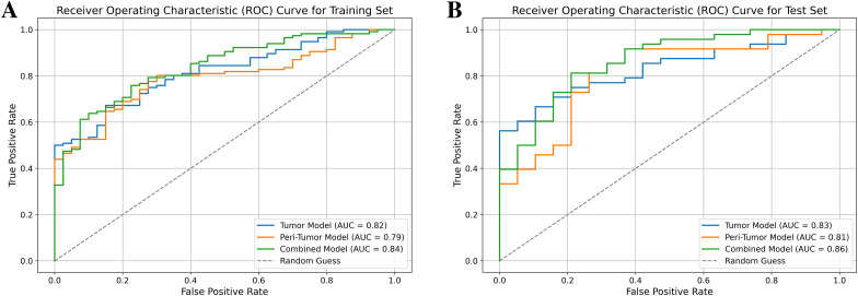
Result: For a Ki-67 cutoff value of 20%, the combined model exhibited the highest performance, with area under the curve (AUC) values of 0.838 (95% confidence interval (CI): 0.774-0.897) for the training set and 0.863 (95% CI: 0.764-0.949) for the test set. The AUC values for the tumor model were 0.816 (95% CI: 0.745-0.880) and 0.830 (95% CI: 0.724-0.916), and for the peritumor model were 0.790 (95% CI: 0.711-0.857) and 0.808 (95% CI: 0.682-0.910). When the Ki-67 cutoff value was set at 29%, the combined model also demonstrated superior predictive ability in both training set (AUC: 0.796; 95% CI: 0.724-0.862) and the test set (AUC: 0.823; 95% CI: 0.723-0.911). The AUC values for the tumor model were 0.785 (95% CI: 0.708-0.861) and 0.784 (95% CI: 0.663-0.882), and for the peritumor model were 0.773 (95% CI: 0.690-0.844) and 0.729 (95% CI: 0.603-0.847).

Conclusion: Radiomics with DCE-MRI can predict Ki-67 expression in invasive ductal breast cancer. Integrating radiomics features from intratumoral and peritumoral regions yields a dependable prognostic model, facilitating pre-surgical detection and treatment decisions. This holds potential for commercial diagnostic tools.

Keywords: DCE-MRI; Invasive ductal breast cancer; Ki-67; machine learning; radiomics.


Conflict of interest statement

Declaration of Conflicting InterestsThe author(s) declared no potential conflicts of interest with respect to the research, authorship, and/or publication of this article.

Figures

Figure 1.
Figure 1.
A flow chart of the patient recruitment process in this study.
Figure 2.
Figure 2.
Workflow of the study. First, the tumor boundary was delineated layer by layer on each image, and 3D segmented tumor images were obtained using 3D Slicer. Subsequently, a morphological operation of dilation was conducted to segment the surrounding tumor up to a radial distance of 4 mm to obtain the peritumoral ROI. Radiomic features were then extracted using the radiomics plug-in in 3D Slicer software. After normalizing the parameters and reducing dimensionality, characteristic parameters were selected and classified using Elastic Net regression. Next, five machine learning algorithms were assessed to predict the Ki-67 expression level, and the best one was selected. Based on the best machine learning algorithm and selected radiomic features from each ROI, three radiomic models were established: the Tumor model, Peri-tumor model, and Combined model. Finally, the discrimination performance of the radiomic models was evaluated using receiver operating characteristic (ROC) analysis.
Figure 3.
Figure 3.
Feature selection using the Elastic Net (take the intratumoral region for example). (A) Variation of Feature Coefficients with Regularization Parameter in Elastic Net. The figure shows the variation of feature coefficients with different regularization parameters (alphas) in Elastic Net regularization. Each line represents the coefficient of a specific feature as alpha changes. The red dashed line indicates the best alpha value selected by grid search. The x-axis is in logarithmic scale to cover a wide range of alpha values. (B) Grid Search Results. The figure illustrates the grid search results for the Elastic Net model, showing how the negative mean squared error changes with different values of the regularization parameter (Alpha) for various L1 ratios. Each line represents the performance for a specific L1 ratio, with the x-axis representing the Alpha values in logarithmic scale.
Figure 4.
Figure 4.
Feature Selection using Elastic Net. The figure displays features selected from tumor (A/a), peri-tumoral (B/b), and combined (C/c) regions at Ki-67 cut-off values of 20% and 29%. Each bar represents a feature, and its length indicates its importance in the predictive model.
Figure 5.
Figure 5.
(A) Receiver operating characteristic (ROC) of three models for predicting Ki-67 status of cut-off value 20% in the training set. (B) ROC of three models in the test set.
Figure 6.
Figure 6.
(A) Receiver operating characteristic (ROC) of three models for predicting Ki-67 status of cut-off value 29% in the training set. (B) ROC of three models in the test set.

References

    1. Dorling L, Carvalho S, Allen J, et al. Breast cancer risk genes - association analysis in more than 113,000 women. New Engl J Med. 2021;384(5):428-439. doi;10.1056/NEJMoa1913948 - DOI - PMC - PubMed
    1. Giaquinto AN, Sung H, Miller KD, et al. Breast cancer statistics, 2022. CA Cancer J Clin. 2022;72(6):524-541. doi:10.3322/caac.21754 - DOI - PubMed
    1. Sharma GN, Dave R, Sanadya J, et al. Various types and management of breast cancer: An overview. J Adv Pharm Technol Res. 2010;1(2):109-126. PMID: 22247839. - PMC - PubMed
    1. de Azambuja E, Cardoso F, de Castro G, et al. Ki-67 as prognostic marker in early breast cancer: a meta-analysis of published studies involving 12,155 patients. Brit J Cancer. 2007;96(10):1504-1513. doi;10.1038/sj.bjc.6603756 - DOI - PMC - PubMed
    1. Davey MG, Hynes SO, Kerin MJ, et al. Ki-67 as a prognostic biomarker in invasive breast cancer. Cancers (Basel). 2021;13(17):4455. doi:10.3390/cancers13174455 - DOI - PMC - PubMed

MeSH terms