Randomized Controlled Trial
. 2025 Apr 3;148(4):1360-1373.
doi: 10.1093/brain/awae334.

Methylphenidate differentially alters corticostriatal connectivity after traumatic brain injury

Affiliations
Randomized Controlled Trial

Methylphenidate differentially alters corticostriatal connectivity after traumatic brain injury

Emma-Jane Mallas et al. Brain. .

Abstract

Traumatic brain injury commonly impairs attention and executive function and disrupts the large-scale brain networks that support these cognitive functions. Abnormalities of functional connectivity are seen in corticostriatal networks, which are associated with executive dysfunction and damage to neuromodulatory catecholaminergic systems caused by head injury. Methylphenidate, a stimulant medication that increases extracellular dopamine and noradrenaline, can improve cognitive function following traumatic brain injury. In this experimental medicine add-on study to a randomized, double-blind, placebo-controlled clinical trial, we test whether administration of methylphenidate alters corticostriatal network function and influences drug response. Forty-three moderate-severe traumatic brain injury patients received 0.3 mg/kg of methylphenidate or placebo twice a day in 2-week blocks. Twenty-eight patients were included in the neuropsychological and functional imaging analysis (four females, mean age 40.9 ± 12.7 years, range 20-65 years) and underwent functional MRI and neuropsychological assessment after each block. 123I-Ioflupane single-photon emission computed tomography dopamine transporter scans were performed, and specific binding ratios were extracted from caudate subdivisions. Functional connectivity and the relationship to cognition were compared between drug and placebo conditions. Methylphenidate increased caudate to anterior cingulate cortex functional connectivity compared with placebo and decreased connectivity from the caudate to the default mode network. Connectivity within the default mode network was also decreased by methylphenidate administration, and there was a significant relationship between caudate functional connectivity and dopamine transporter binding during methylphenidate administration. Methylphenidate significantly improved executive function in traumatic brain injury patients, and this was associated with alterations in the relationship between executive function and right anterior caudate functional connectivity. Functional connectivity is strengthened to brain regions, including the anterior cingulate, that are activated when attention is focused externally. These results show that methylphenidate alters caudate interactions with cortical brain networks involved in executive control. In contrast, caudate functional connectivity reduces to default mode network regions involved in internally focused attention and that deactivate during tasks that require externally focused attention. These results suggest that the beneficial cognitive effects of methylphenidate might be mediated through its impact on the caudate. Methylphenidate differentially influences how the caudate interacts with large-scale functional brain networks that exhibit co-ordinated but distinct patterns of activity required for attentionally demanding tasks.

Keywords: corticostriatal; executive dysfunction; functional connectivity; methylphenidate; traumatic brain injury.


Conflict of interest statement

The authors report no competing interests.

Figures
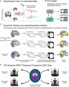
Figure 1
Figure 1
Overview of the methods used to assess the effects of methylphenidate on resting-state functional connectivity in traumatic brain injury. (A) The hypothesized action of methylphenidate (MPH) on caudate connectivity to corticostriatal networks (example time course shown in red, top) and default mode network (DMN, shown in blue, bottom). Under the influence of methylphenidate, caudate connectivity to corticostriatal networks will increase, and caudate connectivity to the DMN will decrease. This will be consistent with a more optimal pattern of functional connectivity for externally directed attention. (B) At baseline, traumatic brain injury (TBI) patients were assessed for functional connectivity abnormalities compared with healthy controls, as reported by De Simoni et al. TBI patients were then randomized into a double-blind, placebo-controlled, crossover clinical trial to investigate the effects of methylphenidate. An MRI scan was performed at the end of each 2-week treatment block. (C) A dual-regression approach was used to evaluate functional connectivity differences between placebo and methylphenidate conditions in TBI patients. Three main analyses were performed to assess cortico-caudate and DMN connectivity. Caudate components and corresponding cortico-caudate networks were defined using the Human Connectome Project resting-state data. The DMN was defined according to Smith et al. (D) 123I-Ioflupane single-photon emission computed tomography (SPECT) dopamine transporter (DAT) scans were performed at baseline as reported by Jenkins et al. Specific binding ratios were extracted from each caudate subdivision to assess the relationship between the degree of DAT binding and changes in functional connectivity with methylphenidate. MNI = Montreal Neurological Institute.
Figure 2
Figure 2
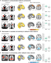
Effects of methylphenidate on resting-state functional connectivity in traumatic brain injury patients. (A) Three caudate subdivisions located in bilateral right and anterior left caudate and associated increases in functional connectivity on methylphenidate (MPH) compared with placebo. For each caudate subdivision, the functional connectivity analysis was constrained to voxels within the corresponding whole-brain network identified within the Human Connectome Project cohort. (B) Anterior and posterior right caudate subdivisions and associated decreases in functional connectivity on MPH compared with placebo. For each caudate subdivision, the functional connectivity analysis was constrained within the default mode network (DMN) defined according to Smith et al. (C) The DMN decreases in functional connectivity on MPH compared to placebo. This functional connectivity analysis was also constrained within the DMN defined according to Smith et al. All results are thresholded at P < 0.05, family-wise error corrected for multiple comparisons. Box plots are presented for visualization purposes only and present functional connectivity values in MPH and placebo conditions extracted from regions demonstrating changes in functional connectivity on MPH. L = left; PLA = placebo; R = right.
Figure 3
Figure 3
The relationship between cortico-caudate functional connectivity and neuropsychological measures. (A) The anterior right caudate subdivision and associated increases in functional connectivity (FC) on methylphenidate (MPH). The FC map is thresholded at P < 0.05, family-wise error corrected for multiple comparisons. (B) Plots representing the relationship between right anterior caudate functional connectivity and performance, including measures of information processing (trail making B) and executive function (Stroop inhibition-switch time versus baseline contrast) corrected for baseline speed. There were significant interactions between treatment condition and cognitive performance (information processing P = 0.026; executive function P = 0.033) on anterior right caudate functional connectivity. Highlighted areas correspond to the 95% confidence intervals. PLA = placebo; R = right; s = seconds.
Figure 4
Figure 4
Relationship of dopamine transporter specific binding ratios and anterior right caudate functional connectivity. (A) A selection of 123I-ioflupane single-photon emission computed tomography dopamine transporter (SPECT DAT) scans of four traumatic brain injury patients. Images from Jenkins et al. The age (years) and sex [all male (M)] of the subject and the time since injury (in months) are shown. The colour scale shows the 123I-ioflupane specific binding ratio (SBR). (B) Voxel-wise reductions in 123I-ioflupane are shown in red/yellow. The striatal mask is shown for comparison in blue. The colour bar shows the corrected P-values. Image from Jenkins et al. (C) Relationship of 123I-ioflupane SBR within the right (R) anterior caudate to functional connectivity (FC) of the right anterior caudate in both placebo (PLA, top) and methylphenidate (MPH, bottom) conditions. This relationship is significant in the methylphenidate condition (P = 0.01). Highlighted areas correspond to the 95% confidence intervals.

References

    1. Draper K, Ponsford J. Cognitive functioning ten years following traumatic brain injury and rehabilitation. Neuropsychology. 2008;22:618–625. - PubMed
    1. Pavlovic D, Pekic S, Stojanovic M, Popovic V. Traumatic brain injury: Neuropathological, neurocognitive and neurobehavioral sequelae. Pituitary. 2019;22:270–282. - PubMed
    1. Little DM, Kraus MF, Joseph J, et al. Thalamic integrity underlies executive dysfunction in traumatic brain injury. Neurology. 2010;74:558–564. - PMC - PubMed
    1. Leunissen I, Coxon JP, Caeyenberghs K, Michiels K, Sunaert S, Swinnen SP. Task switching in traumatic brain injury relates to cortico-subcortical integrity. Hum Brain Mapp. 2014;35:2459–2469. - PMC - PubMed
    1. Peters SK, Dunlop K, Downar J. Cortico-striatal-thalamic loop circuits of the salience network: A central pathway in psychiatric disease and treatment. Front Syst Neurosci. 2016;10:104. - PMC - PubMed

Publication types

MeSH terms

Substances