. 2024 Dec:90:102050.
doi: 10.1016/j.molmet.2024.102050. Epub 2024 Oct 20.

Interruption of glucagon signaling augments islet non-alpha cell proliferation in SLC7A2- and mTOR-dependent manners

Affiliations

Interruption of glucagon signaling augments islet non-alpha cell proliferation in SLC7A2- and mTOR-dependent manners

Katie C Coate et al. Mol Metab. 2024 Dec.

Abstract

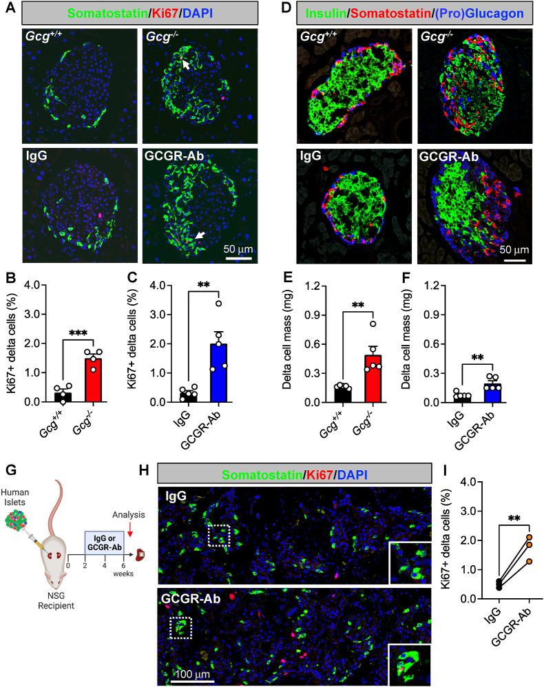
Objective: Dysregulated glucagon secretion and inadequate functional beta cell mass are hallmark features of diabetes. While glucagon receptor (GCGR) antagonism ameliorates hyperglycemia and elicits beta cell regeneration in pre-clinical models of diabetes, it also promotes alpha and delta cell hyperplasia. We sought to investigate the mechanism by which loss of glucagon action impacts pancreatic islet non-alpha cells, and the relevance of these observations in a human islet context.

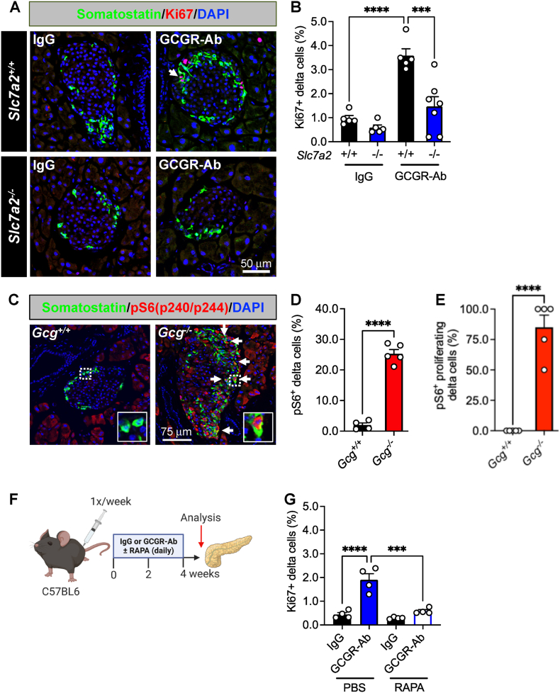
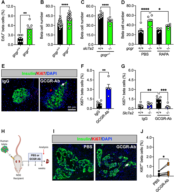
Methods: We used zebrafish, rodents, and transplanted human islets comprising six different models of interrupted glucagon signaling to examine their impact on delta and beta cell proliferation and mass. We also used models with global deficiency of the cationic amino acid transporter, SLC7A2, and mTORC1 inhibition via rapamycin, to determine whether amino acid-dependent nutrient sensing was required for islet non-alpha cell growth.

Results: Inhibition of glucagon signaling stimulated delta cell proliferation in mouse and transplanted human islets, and in mouse islets. This was rapamycin-sensitive and required SLC7A2. Likewise, gcgr deficiency augmented beta cell proliferation via SLC7A2- and mTORC1-dependent mechanisms in zebrafish and promoted cell cycle engagement in rodent beta cells but was insufficient to drive a significant increase in beta cell mass in mice.

Conclusions: Our findings demonstrate that interruption of glucagon signaling augments islet non-alpha cell proliferation in zebrafish, rodents, and transplanted human islets in a manner requiring SLC7A2 and mTORC1 activation. An increase in delta cell mass may be leveraged for future beta cell regeneration therapies relying upon delta cell reprogramming.

Keywords: Beta cell; Delta cell; Glucagon receptor; Pancreatic islet; Proliferation; mTOR.


Conflict of interest statement

Declaration of competing interest Hai Yan is an employee of REMD Biotherapeutics Inc. which licensed one of the glucagon receptor antibodies used for some of the studies in this manuscript. All other authors have no interests to declare.

Figures

Figure 1
Figure 1
Loss of glucagon action augments delta cell proliferation and mass expansion in mouse and transplanted human islets. (A) Representative images of pancreatic islet and delta cell proliferation in 8 week old Gcg+/+/Gcg−/− (upper row) and 2 week treated IgG/GCGR-Ab-treated C57BL6 (bottom row) mice. Somatostatin (green), Ki67 (red), and DAPI (blue) are shown. White arrows indicate Ki67+ somatostatin + cells. (B, C) Quantification of pancreatic islet delta cell proliferation in (B) Gcg+/+ (black bar) and Gcg−/− (red bar) mice (n = 1–2 females and 2–3 males per genotype) and (C) control IgG (black bar) and GCGR-Ab-treated (blue bar) mice (all males, unpaired t test, ∗∗∗p < 0.001 versus Gcg+/+, ∗∗p < 0.01 versus IgG). (D) Representative images of pancreatic islet hormones in Gcg+/+/Gcg−/− (upper row) and IgG/GCGR-Ab-treated (bottom row) mice. Insulin (green), somatostatin (red), and pro-glucagon (Gcg+/+/Gcg−/−; blue) or glucagon (IgG/GCGR-Ab; blue) are shown. (E, F) Pancreatic islet delta cell mass in (E) Gcg+/+ (black bar) and Gcg−/− (red bar) mice (n = 1–2 females and 3 males per genotype) and (F) control IgG (black bar) and GCGR-Ab-treated (blue bar) mice (all males, unpaired t test, ∗∗p < 0.01 versus Gcg+/+ or IgG). (G) Schematic of approach for human islet subcapsular renal transplantation in NSG recipient mice followed by control IgG or GCGR-Ab treatment. Created with BioRender.com (H) Representative images of delta cell proliferation in human islet grafts after 4 weeks of control IgG (upper row) or GCGR-Ab-treatment (bottom row). Grafts were immunostained for somatostatin (green), Ki67 (red), and DAPI (blue). White dashed boxes indicate regions selected for insets. (I) Quantification of delta cell proliferation in transplanted human islets in control IgG (black circles) and GCGR-Ab-treated (orange circles) mice (n = 3 donors [see Supplemental Table 1], unpaired t test, ∗∗p < 0.01 versus IgG).
Figure 2
Figure 2
SLC7A2 and mTOR activation are required for delta cell proliferation in response to interrupted glucagon signaling. (A) Representative images of pancreatic islet delta cell proliferation in Slc7a2+/+ (upper row) and Slc7a2−/− (bottom row) IgG/GCGR-Ab-treated mice. Somatostatin (green), Ki67 (red), and DAPI (blue) are shown. White arrows indicate Ki67+ somatostatin + cells. (B) Quantification of pancreatic islet delta cell proliferation in Slc7a2+/+ (black bars) and Slc7a2−/− (blue bars) IgG or GCGR-Ab-treated mice (n = 4 females and 1–3 males per genotype, one-way ANOVA with Tukey's multiple comparisons test, ∗∗∗∗p < 0.0001 versus Slc7a2+/+ IgG, ∗∗∗p < 0.001 versus Slc7a2+/+ GCGR-Ab). (C) Representative images of pancreatic islets in Gcg+/+/Gcg−/− mice immunostained for somatostatin (green), phosphorylated ribosomal protein S6 (pS6240/244; red), Ki67 (white), and DAPI (blue). White arrows indicate pS6+ somatostatin + cells. Yellow arrows indicate pS6+ Ki67+ somatostatin + cells. White dashed boxes indicate regions selected for insets. (D) Quantification of the percentage of pS6+ somatostatin + cells in Gcg+/+ (black bar) and Gcg−/− (red bar) pancreatic islets (n = 1–2 females and 3–4 males per genotype, unpaired t test, ∗∗∗∗p < 0.0001 versus Gcg+/+). (E) Quantification of the percentage of pKi67+ somatostatin + cells that are also pS6+ in Gcg+/+ (black bar) and Gcg−/− (red bar) pancreatic islets (n = 3–4 females and 3 males per genotype, unpaired t test, ∗∗∗∗p < 0.0001 versus Gcg+/+). (F) Schematic of approach for IgG/GCGR-Ab (once weekly) and rapamycin (RAPA; once daily) co-treatment in C57BL6 mice. Created with BioRender.com (G) Quantification of pancreatic islet delta cell proliferation in mice co-treated with IgG (black bars) or GCGR-Ab (blue and white bar) and PBS or RAPA (all males, one-way ANOVA with Tukey's multiple comparisons test, ∗∗∗∗p < 0.0001 versus IgG, ∗∗∗p < 0.001 versus PBS/GCGR-Ab).
Figure 3
Figure 3
Loss of glucagon receptor function stimulates beta cell proliferation in a species-specific manner. (A) Beta cells stained for EdU to assess their proliferation in 5 dpf (days post-fertilization) wild-type (gcgr+/+, black bar) and gcgra/b−/− (abbreviated gcgr−/−, green bar) zebrafish (n = 8 per group, unpaired t test, ∗∗p < 0.01 versus gcgr+/+). (B) Beta cell number in 5 dpf gcgr+/+ (black bar) and gcgr−/− (green bar) zebrafish (n = 24–30 per group, unpaired t test, ∗∗∗∗p < 0.0001 versus gcgr+/+). (C) Beta cell number after knockdown of slc7a2 (+/+, black bar; −/−, green bar) in 5 dpf gcgr−/− zebrafish (n = 8–15 per group, unpaired t test, ∗∗∗∗p < 0.0001 versus slc7a2+/+). (D) Beta cell number in 6 dpf gcgr+/+ (black bar) and gcgr−/− (green bar) zebrafish after 3 days of treatment with PBS or RAPA (n = 6–7 per group, one-way ANOVA with Fisher's LSD, ∗∗∗∗p < 0.0001 versus PBS/gcgr+/+, ∗p < 0.05 versus PBS/gcgr−/−). (E) Representative images of pancreatic islet beta cell proliferation in 8 week treated IgG/GCGR-Ab-treated C57BL6 mice. Insulin (green), Ki67 (red), and DAPI (blue) are shown. White arrow indicates a Ki67+ insulin + cell. (F) Quantification of pancreatic islet beta cell proliferation in control IgG (black bar) and GCGR-Ab-treated (blue bar) mice (all males, unpaired t test, ∗∗p < 0.01 versus IgG). (G) Quantification of pancreatic islet beta cell proliferation in Slc7a2+/+ (black bars) and Slc7a2−/− (blue bars) IgG or GCGR-Ab-treated mice (n = 2–5 females and 3–6 males per group, one way ANOVA with Tukey's multiple comparisons test, ∗∗p < 0.0001 versus Slc7a2+/+ IgG, ∗∗∗p < 0.001 versus Slc7a2+/+ GCGR-Ab). (H) Schematic of approach for human islet subcapsular renal transplantation in NSG recipient mice followed by PBS or GCGR-Ab treatment. Created with BioRender.com (I) Representative images of beta cell proliferation in human islet grafts after 4 weeks of PBS or GCGR-Ab-treatment. Grafts were immunostained for insulin (green), Ki67 (red), and DAPI (blue). Dashed yellow lines indicate kidney-graft boundary. (J) Quantification of beta cell proliferation in transplanted human islets in PBS (black circles) or GCGR-Ab-treated (orange circles) mice (n = 2 female and 5 male donors [see Supplemental Table 1], unpaired t test, ∗p < 0.05 versus PBS).

Update of

References

    1. Muller W.A., Faloona G.R., Aguilar-Parada E., Unger R.H. Abnormal alpha-cell function in diabetes. Response to carbohydrate and protein ingestion. N Engl J Med. 1970;283(3):109–115. - PubMed
    1. Unger R., Orci L. The essential role of glucagon in the pathogenesis of diabetes mellitus. Lancet. 1975;305(7897):14–16. - PubMed
    1. Kahn S.E. The importance of β-cell failure in the development and progression of type 2 diabetes. J Clin Endocrinol Metabol. 2001;86(9):4047–4058. - PubMed
    1. Dunning B.E., Gerich J.E. The role of alpha-cell dysregulation in fasting and postprandial hyperglycemia in type 2 diabetes and therapeutic implications. Endocr Rev. 2007;28(3):253–283. - PubMed
    1. Hudish L.I., Reusch J.E.B., Sussel L. β Cell dysfunction during progression of metabolic syndrome to type 2 diabetes. J Clin Investig. 2019;129(10):4001–4008. - PMC - PubMed

MeSH terms

Substances

LinkOut - more resources