. 2024 Oct 21;10(1):444.
doi: 10.1038/s41420-024-02211-z.

Upregulation of cholinergic modulators Lypd6 and Lypd6b associated with autism drives anxiety and cognitive decline

Affiliations

Upregulation of cholinergic modulators Lypd6 and Lypd6b associated with autism drives anxiety and cognitive decline

Aizek B Isaev et al. Cell Death Discov. .

Abstract

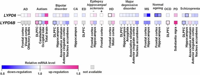
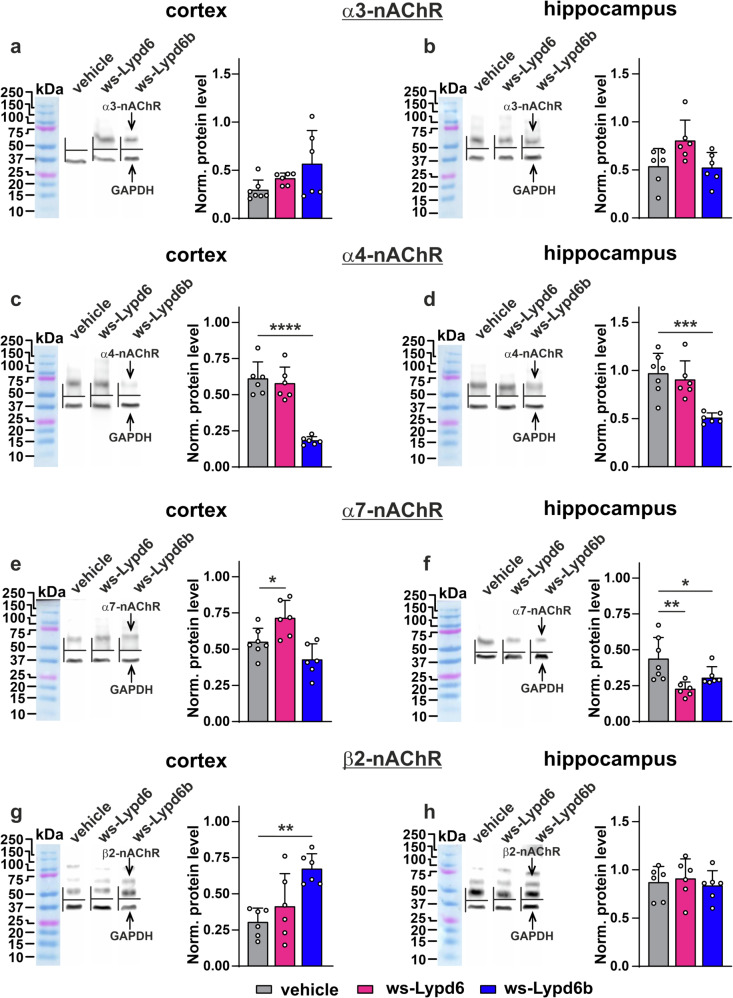
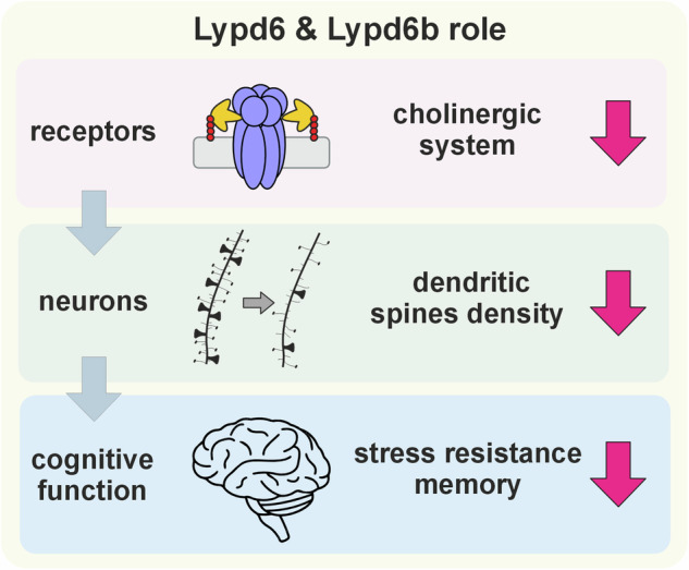
Intellectual disability and autistic features are associated with chromosome region 2q23.q23.2 duplication carrying LYPD6 and LYPD6B genes. Here, we analyzed LYPD6 and LYPD6B expression in patients with different neuropsychiatric disorders. Increased LYPD6 and LYPD6B expression was revealed in autism and other disorders. To study possible consequences of Lypd6 and Lypd6b overexpression in the brain, we used a mouse model with intracerebroventricular delivery of recombinant analogs of these proteins. A two-week infusion evoked significant memory impairment and acute stress. Both modulators downregulated hippocampal and amygdala dendritic spine density. No changes in synaptic plasticity were observed. Intracerebroventricular administration by both proteins downregulated hippocampal expression of Lypd6, Lypd6b, and α7 nicotinic acetylcholine receptor (nAChR). Similar to Lypd6, Lypd6b targeted different nAChR subtypes in the brain with preferential inhibition of α7- and α4β2-nAChRs. Thus, increased Lypd6 and Lypd6b level in the brain are linked to cholinergic system depression, neuronal atrophy, memory decline, and anxiety.


Conflict of interest statement

The authors declare no competing interests.

Figures

Fig. 1
Fig. 1. LYPD6 and LYPD6B mRNA expression in the brain of patients with neurological and neuropsychiatric disorders.
Data presented as relative mRNA level in diseased patients as compared to healthy donors. For the dataset accession numbers, number of patients and statistical details, see Supplementary Table 1. AD Alzheimer’s disease, CA chronic alcoholism, DLPFC dorsolateral prefrontal cortex, ED eating disorder, HD Huntington’s disease, MS multiple sclerosis, OCR Obsessive-compulsive disorder, PD Parkinson’s disease. For normal ageing, people younger 35 y.o. and older than 85 y.o. were compared. Boxes with the thick outlines represent significant difference between healthy donors and patients with neurological and neuropsychiatric disorders according to two-sided t-test. p values are in Supplementary Table 1.
Fig. 2
Fig. 2. Working memory impairment and stress induction upon mice administration with ws-Lypd6 and ws-Lypd6b.
ae Open field test. Data presented as the value of an experimentally estimated parameter ± SEM (n = 1720). *(p < 0.05) indicates significant difference from vehicle group according to Kruskal–Wallis test followed by post hoc Dunn’s test. f Rotarod test over the 7 consecutive days expressed as retention time on the accelerating rotarod roller (n = 17–19). No difference between the groups according to two-way ANOVA followed by post hoc Dunnet test. gi Elevated plus maze. Data presented as the value of an experimentally estimated parameter ± SEM (n = 15–19). No difference between groups according to Kruskal–Wallis test followed by post hoc Dunn’s test. j Acute stress hypophagia test. Consumption of sweetened milk under comfortable conditions and upon acute stress in a novel environment. Data presented as sweetened milk consumption (ml) ± SEM (n = 17–20). *(p < 0.05) and **(p < 0.01) indicate significant difference between data groups according to two-way ANOVA with post hoc Sidak test. k, l Novel object and novel odor recognition tests. Data presented as preference index ± SEM (n = 17–20). ##(p < 0.01) and ###(p < 0.001) indicate significant difference in preference of novel object/novel odor over a familiar one according to one-sample Wilcoxon-test. *(p < 0.05) indicates significant difference between data groups according to Kruskal–Wallis test followed by post hoc Dunn’s test.
Fig. 3
Fig. 3. Decreased dendritic spine density in the amygdala and hippocampus following ws-Lypd6 and ws-Lypd6b administration in mice.
ac Left panels. Representative images of dendrites with spines of neurons from the cortex, amygdala, and hippocampus of experimental mice. ac Right panels. Number of stub and mushroom-shaped dendritic spines in the cortex, amygdala, and hippocampus of treated mice. Data presented as a number of dendritic spines of different types (per 10 µm) ± SEM (n = 34). **(p < 0.01) and ****(p < 0.0001) indicate significant difference from vehicle group according to two-way ANOVA test following post hoc Dunnet test.
Fig. 4
Fig. 4. Ws-Lypd6 and ws-Lypd6b administration does not influence hippocampal synaptic plasticity.
a Averaged normalized field excitatory postsynaptic potentials (fEPSPs) in hippocampal slices after drug administration were recorded for 1 h (nws-Lypd6 = 9, nws-Lypd6b = 8, nvehicle = 7) after HFS. Representative traces are shown above: black – baseline, colored – post-tetanic recording. b Normalized fEPSP slopes averaged over 0–10 and 50–60 min after HFS in the hippocampal slices. c PPF ratio upon stabilization of baseline fEPSP responses. Representative fEPSP traces for 50 ms interpulse interval stimulus are shown above. No difference between groups according to two-way ANOVA followed by post hoc Dunnett’s test (a, c) or one-way ANOVA followed by post hoc Dunnet test (b). dh genes and brain regions that showed statistically significant differences in expression compared to the control group. Data are presented as the ratio of gene expression level ± SEM (n = 4–12). *(p < 0.05) and **(p < 0.01) indicate difference from control according to one-way ANOVA test followed by post hoc Dunnett’s test.
Fig. 5
Fig. 5. Changes in expression of α3-, α4-, α7-, and β2-nAChR subunits in the cortex and hippocampus upon ws-Lypd6 and ws-Lypd6b administration.
ah Left panels. Representative western blot bands for analysis of expression of α3-, α4-, α7-, and β2- nAChR subunits in the cortex (a, c, e, g) and hippocampus (b, d, f, h). ah Right panels. Quantification of the α3-, α4-, α7-, and β2- nAChR subunits expression level normalized to GAPDH expression level. Data presented as normalized protein band intensity ± SEM (n = 6–7). *(p < 0.05), **(p < 0.01), and ****(p < 0.0001) indicate significant difference from vehicle group according to two-way ANOVA following post hoc Dunnett’s test. Whole western blotting membranes are in Supplementary Figs. 4 and 5.
Fig. 6
Fig. 6. Changes in expression of endogenous Lypd6 and Lypd6b in the cortex and hippocampus upon ws-Lypd6 and ws-Lypd6b administration.
ad Left panels. Representative western blot bands for analysis of expression of endogenous Lypd6 and Lypd6b in the cortex (a, c) and hippocampus (b, d). ad Right panels. Quantification of the Lypd6, Lypd6b expression level normalized to β-actin expression level. Data presented as normalized protein band intensity ± SEM (n = 6–7). *(p < 0.05), **(p < 0.01), and ****(p < 0.0001) indicate significant difference from vehicle group according to two-way ANOVA following post hoc Dunnett’s test. Whole western blotting membranes are in Supplementary Fig. 6.
Fig. 7
Fig. 7. Ws-Lypd6b inhibits multiple nAChRs and binds subunits of different nicotinic and GABAA receptors.
a Normalized representative responses to 100 ms pulses of ACh (100 µM for α7, α3β2, α3β4, α4β2 LS, 10 µM for α4β2 HS) recorded at different nAChRs expressed in X. laevis oocytes in presence or absence of 30 μM ws-Lypd6b. Pre-incubation time of oocytes with ws-Lypd6b was 20 s. b Effect of 30 μM ws-Lypd6b on the ACh-evoked current at different nAChRs. Data presented as normalized ACh-evoked currents in the presence or the absence of ws-Lypd6b ± SEM (n = 5–13 oocytes). **(p < 0.01) indicates significant difference from control (100%, dashed line) according to one-sample Wilcoxon test following post hoc Holm-Sidak test. c. Dose-response curves for inhibition of ACh-evoked currents at different nAChRs by ws-Lypd6b. Data normalized to peak current amplitude recorded without ws-Lypd6b (100%), presented as mean ± SEM (n = 5–10 oocytes, each of them was treated by 0.5–100 µM ws-Lypd6b) and fit Hill’s equation. Please note that the difference in data between b and c is because experiments were carried out independently. d Analysis of the receptor subunits extracted by ws-Lypd6b from the total brain homogenate (n = 3–4). Empty NHS-Sepharose blocked with 500 mM ethanolamine was used as control. Whole western-blotting membranes are in Supplementary Fig. 7.
Fig. 8
Fig. 8. General proposed mechanism of Lypd6 and Lypd6b action in the CNS.
Increased abundance of Lypd6 and Lypd6b in the brain impairs cognitive function at different physiological levels from neuronal receptors to neurons and behavior.

References

    1. Terry AV, Jones K, Bertrand D. Nicotinic acetylcholine receptors in neurological and psychiatric diseases. Pharmacol Res. 2023;191:106764. - PubMed
    1. Mineur YS, Soares AR, Etherington IM, Abdulla ZI, Picciotto MR. Pathophysiology of nAChRs: limbic circuits and related disorders. Pharmacol Res. 2023;191:106745. - PubMed
    1. Picciotto MR, Lewis AS, van Schalkwyk GI, Mineur YS. Mood and anxiety regulation by nicotinic acetylcholine receptors: a potential pathway to modulate aggression and related behavioral states. Neuropharmacology. 2015;96:235–43. - PMC - PubMed
    1. Thomsen MS, Weyn A, Mikkelsen JD. Hippocampal α7 nicotinic acetylcholine receptor levels in patients with schizophrenia, bipolar disorder, or major depressive disorder. Bipolar Disord. 2011;13:701–7. - PubMed
    1. Hannestad JO, Cosgrove KP, DellaGioia NF, Perkins E, Bois F, Bhagwagar Z, et al. Changes in the cholinergic system between bipolar depression and euthymia as measured with [123I]5IA single photon emission computed tomography. Biol Psychiatry. 2013;74:768–76. - PMC - PubMed

LinkOut - more resources