. 2024 Sep 19;27(10):110993.
doi: 10.1016/j.isci.2024.110993. eCollection 2024 Oct 18.

Regorafenib promotes antitumor progression in melanoma by reducing RRM2

Affiliations

Regorafenib promotes antitumor progression in melanoma by reducing RRM2

Xiuyun Xuan et al. iScience. .

Abstract

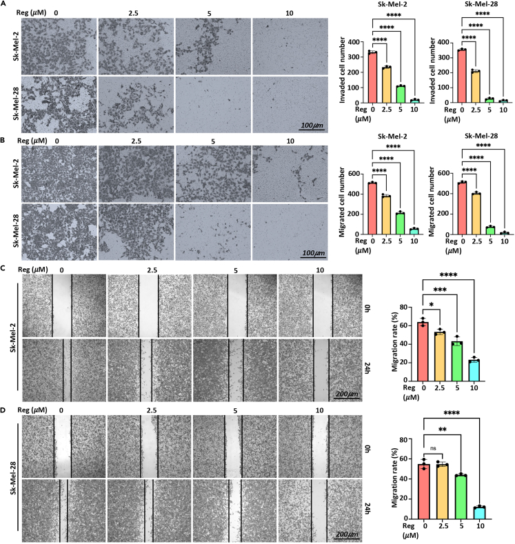
Melanoma is a malignant tumor with a terrible prognosis. Although so many therapies are used for melanoma, the overall survival rate is still poor globally. Novel therapies are still required. In our study, the role and potential mechanism of regorafenib in melanoma are explored. Regorafenib has the ability to limit the growth, invasion, and metastasis of melanoma cells but to upregulate apoptosis-prompting markers (cleaved-PARP and Bax). RRM2 is identified to be the downstream target of regorafenib by RNA sequencing. In addition, we discovered that RRM2 inhibition and regorafenib have comparable effects on melanoma cells. Rescue experiments showed that RRM2 is crucial in regulating regorafenib's anti-melanoma progression. Moreover, ERK/E2F3 signaling influences regorafenib's ability to suppress melanoma cell growth. Ultimately, regorafenib significantly inhibits tumor growth in vivo. In conclusion, our finding demonstrated that regorafenib promotes antitumor progression in melanoma by reducing RRM2.

Keywords: Biological sciences; Cancer; Therapy.


Conflict of interest statement

The authors declare no competing interests.

Figures

None
Graphical abstract
Figure 1
Figure 1
Regorafenib significantly inhibited melanoma cells growth (A) Regorafenib had an obvious cytotoxicity on melanoma cells in a concentration- and time-dependent manner. (B–G) Regorafenib markedly inhibited melanoma cells growth by cell colony formation (B–D) and EDU staining (E–G. Scale bar: 100 μm). (H) Dose-dependent regorafenib treatment blocked the cell cycle transition from G1 to S in Sk-Mel-2/28 cell lines. (I) The levels of CDK4 and cyclin D1, G1 phase-regulated proteins, were reduced by regorafenib in Sk-Mel-2/28 cell lines. Reg: regorafenib. ∗p < 0.05, ∗∗p < 0.01, ∗∗∗p < 0.001, ∗∗∗∗p < 0.0001. Data are represented as mean ± SEM. Similar results were obtained in two additional independent experiments. Data are represented as mean ± SEM.
Figure 2
Figure 2
Regorafenib suppressed the invasive and migrated capacities of melanoma cells (A) Regorafenib reduced the proportion of invaded cells. (B–D) Regorafenib significantly decreased the quantity of migrating cells and inhibited melanoma cell wound repair. Reg: regorafenib. ∗p < 0.05, ∗∗p < 0.01, ∗∗∗p < 0.001, ∗∗∗∗p < 0.0001. Data are represented as mean ± SEM. Similar results were obtained in two additional independent experiments. (A + B) Scale bar: 100 μm. (C + D) Scale bar: 200 μm.
Figure 3
Figure 3
Regorafenib induced apoptosis in melanoma cells (A and B) The expression of Bax and cleaved-PARP were increased, while the expression of Bcl-2 was decreased after melanoma cells were treated with regorafenib for 48 h, which were confirmed by western blot analysis (A) and immunofluorescence staining (B, Scale bar: 50 μm). (C) Regorafenib increased the frequencies of apoptotic cells by FCM. Reg: regorafenib. ∗∗p < 0.01, ∗∗∗∗p < 0.0001. Data are represented as mean ± SEM. Similar results were obtained in two additional independent experiments.
Figure 4
Figure 4
RRM2 was the downstream target of regorafenib (A and B) PCA suggested a clear distinction between regorafenib treatment and control samples. (C) 1502 upregulated and 780 downregulated DEGs were identified in regorafenib treatment samples by RNA-seq. (D and E) Downregulated DEGs were used for enrichment analysis. (F) 25 ODEGs were identified through combining analysis of our RNA-seq_down genes and GSE7553_up genes. (G) The levels of RRM2 protein were suppressed when melanoma cells were treated with different concentrations of regorafenib. (H) The predicted binding mode of regorafenib to RRM2 by docking analysis. Reg: regorafenib. ∗∗p < 0.01, ∗∗∗p < 0.001, ∗∗∗∗p < 0.0001. Data are represented as mean ± SEM. Similar results were obtained in two additional independent experiments.
Figure 5
Figure 5
Depletion of RRM2 alleviated the malignancy of melanoma cells (A) The depletion efficiency of RRM2 was confirmed by western blot analysis. (B–E) Depletion of RRM2 significantly inhibited the proliferation of Sk-Mel-2/28 cells by CCK8 (B) and EDU staining (C–E). (F–I) RRM2 depletion reduced the number of invaded cells (F–H) and retarded the wound closure of melanoma cells (I). ∗p < 0.05, ∗∗p < 0.01, ∗∗∗p < 0.001, ∗∗∗∗p < 0.0001. Data are represented as mean ± SEM. Similar results were obtained in two additional independent experiments. (C + F) Scale bar: 100 μm.
Figure 6
Figure 6
RRM2 overexpression partially reversed regorafenib-induced growth inhibition of melanoma cells (A) RRM2 overexpression partially reversed regorafenib-induced cytotoxicity of melanoma cells by CCK8 cytotoxicity test. (B and C) RRM2 overexpression partially reduced regorafenib-induced apoptosis of melanoma cells by FCM. (D and E) RRM2 overexpression partially reverses the suppression of invasion and migration induced by regorafenib in melanoma cells by transell assays. Scale bar: 100 μm. Reg: regorafenib. ∗∗∗p < 0.001, ∗∗∗∗p < 0.0001. Data are represented as mean ± SEM. Similar results were obtained in two additional independent experiments.
Figure 7
Figure 7
ERK/E2F3 signaling participated the inhibition of regorafenib on melanoma cells (A) The expression of p-ERK and E2F3 were inhibited when melanoma cells were treated with regorafenib by western blot. (B) When ERK signaling was blocked, the level of p-ERK, E2F3, and RRM2 was reduced. (C) When E2F3 expression was depleted, RRM2 expression was also depleted. Reg: regorafenib. ∗p < 0.05, ∗∗p < 0.01, ∗∗∗p < 0.001, ∗∗∗∗p < 0.0001. Data are represented as mean ± SEM. Similar results were obtained in two additional independent experiments.
Figure 8
Figure 8
Regorafenib reduced tumorigenesis in vivo (A–C) Regorafenib inhibited the growth of xenograft tumors, including tumor weight (B) and volume (C). (D) Regorafenib treatment inhibited the levels of p-ERK and RRM2, while RRM2 overexpression partially relieved the inhibition of p-ERK and RRM2 expression. (E) There were no obvious differences in the weight of these vital organs between control and regorafenib-treatment group. ∗∗p < 0.01, ∗∗∗p < 0.001, ∗∗∗∗p < 0.0001. Data are represented as mean ± SEM. Similar results were obtained in two additional independent experiments.

References

    1. Carr S., Smith C., Wernberg J. Epidemiology and Risk Factors of Melanoma. Surg. Clin. 2020;100:1–12. doi: 10.1016/j.suc.2019.09.005. - DOI - PubMed
    1. Davis L.E., Shalin S.C., Tackett A.J. Current state of melanoma diagnosis and treatment. Cancer Biol. Ther. 2019;20:1366–1379. doi: 10.1080/15384047.2019.1640032. - DOI - PMC - PubMed
    1. Zeng H., Li J., Hou K., Wu Y., Chen H., Ning Z. Melanoma and Nanotechnology-Based Treatment. Front. Oncol. 2022;12 doi: 10.3389/fonc.2022.858185. - DOI - PMC - PubMed
    1. Swetter S.M., Tsao H., Bichakjian C.K., Curiel-Lewandrowski C., Elder D.E., Gershenwald J.E., Guild V., Grant-Kels J.M., Halpern A.C., Johnson T.M., et al. Guidelines of care for the management of primary cutaneous melanoma. J. Am. Acad. Dermatol. 2019;80:208–250. doi: 10.1016/j.jaad.2018.08.055. - DOI - PubMed
    1. Castet F., Garcia-Mulero S., Sanz-Pamplona R., Cuellar A., Casanovas O., Caminal J.M., Piulats J.M. Uveal Melanoma, Angiogenesis and Immunotherapy, Is There Any Hope? Cancers. 2019;11 doi: 10.3390/cancers11060834. - DOI - PMC - PubMed

LinkOut - more resources