Cavernous Hemangioma of the Tongue Base: A Rare Case
- PMID: 39435252
- PMCID: PMC11492155
- DOI: 10.7759/cureus.69759
Cavernous Hemangioma of the Tongue Base: A Rare Case
Abstract
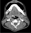
Vascular anomalies encompass a range of conditions affecting blood vessel development, categorized as tumors or malformations. Hemangiomas, the most common vascular tumors, involve abnormal endothelial cell proliferation, particularly in hemangiomas, which are prevalent benign tumors arising from mesenchymal tissue in the head and neck. They manifest as capillary, cavernous, or mixed types, affecting areas like the tongue and lips. Hemangiomas of the tongue base are notably rare, emphasizing the complexity of diagnosis and management due to their uncommon occurrence and potential for complications like bleeding. This report highlights a case of cavernous hemangioma of the tongue base, underscoring diagnostic challenges and management considerations. A Malay man in his late 30s, a nonsmoker and nondrinker, presented with a year-long history of intermittent globus sensation without associated symptoms like odynophagia, dysphagia, intraoral bleeding, or neck swelling. Flexible nasopharyngolaryngoscopy revealed a lobulated bluish mass at the right base of the tongue, prompting a provisional diagnosis of hemangioma. Contrast-enhanced CT suggested an irregular lesion with calcification, leading to MRI confirmation of a well-defined, non-muscle-invasive lesion that favored venolymphatic malformation rather than hemangioma. However, it was confirmed histologically as cavernous hemangioma after excision, where intraoperative findings aligned with initial preoperative clinical assessments.
Keywords: base of the tongue; cavernous hemangioma; head and neck neoplasms; hemangioma; vascular anomalies.
Copyright © 2024, Mohamed et al.
Conflict of interest statement
Human subjects: Consent was obtained or waived by all participants in this study. Conflicts of interest: In compliance with the ICMJE uniform disclosure form, all authors declare the following: Payment/services info: All authors have declared that no financial support was received from any organization for the submitted work. Financial relationships: All authors have declared that they have no financial relationships at present or within the previous three years with any organizations that might have an interest in the submitted work. Other relationships: All authors have declared that there are no other relationships or activities that could appear to have influenced the submitted work.
Figures


References
-
- Vascular anomalies classification: recommendations from the International Society for the study of vascular anomalies. Wassef M, Blei F, Adams D, et al. Pediatrics. 2015;136:203–214. - PubMed
-
- An evaluation of the efficacy of a topical gel with triester glycerol oxide (TGO) in the treatment of minor recurrent aphthous stomatitis in a Turkish cohort: a randomized, double-blind, placebo-controlled clinical trial. [ Oct; 2024 ];Ofluoglu D, Ergun S, Warnakulasuriya S, Namdar-Pekiner F, Tanyeri H. https://doi.org/10.4317/medoral.21469 Med Oral Patol Oral Cir Bucal. 2017 22:159–166. - PMC - PubMed
-
- Current concepts related to the diagnosis and treatment of hemangioma: literature review . de Oliveira Campos IJ, de Medeiros ER, da Costa EH, de Paula JV, Xavier LG, Medeiros CK, da Cruz Lima JG. Res Soc. 2021;10:45510112001.
-
- Hemangioma of base of tongue. Qureshi SS, Chaukar DA, Pathak KA, Sanghvi VD, Sheth T, Merchant NH, Dcruz AK. https://pubmed.ncbi.nlm.nih.gov/15659874/ Indian J Cancer. 2004;41:181–183. - PubMed
-
- Cavernous hemangioma of the tongue: a rare case report. Kamala KA, Ashok L, Sujatha GP. https://www.ncbi.nlm.nih.gov/pmc/articles/PMC4012128/ Contemp Clin Dent. 2014;5:95–98. - PMC - PubMed
Publication types
LinkOut - more resources
Full Text Sources
