. 2024 Oct 7:14:1421443.
doi: 10.3389/fonc.2024.1421443. eCollection 2024.

RUNX2 as a novel biomarker for early identification of patients progressing to advanced-stage mycosis fungoides

Affiliations

RUNX2 as a novel biomarker for early identification of patients progressing to advanced-stage mycosis fungoides

Maria Danielsen et al. Front Oncol. .

Abstract

Introduction: The majority of patients with mycosis fungoides (MF) have an indolent disease course, but a substantial fraction (20-30%) of patients progress to advanced stages - usually with a grave prognosis. Early differentiation between indolent and aggressive types of MF is important for the choice of treatment regimen and monitoring of the individual patient. Good biomarkers are therefore desired.

Methods: Here, we used spatial transcriptomics on skin samples at time-of-diagnosis to enable prediction of patients who later progressed to advanced stages of MF. Formalin-fixed, paraffin-embedded skin biopsies at time of diagnosis from six patients with MF who progressed to advanced stages of disease within 4 months to 12 years after diagnosis, and nine patients who remained in early-stage disease over 9 to 27 years were analyzed using the GeoMx Digital Spatial Profiler to capture spatially resolved high-plex RNA gene expression data. Five different regions of interest (the epidermis, the basal layer of epidermis, CD4+ T-cells and neighboring cells, and Pautrier's microabscesses) were profiled for further assessment.

Results and discussion: Interestingly, RUNX2, SHMT2, and MCM7 were upregulated in the enriched population of malignant T-cells in Pautrier's microabscesses in patients who later developed advanced stages of disease. Expression of RUNX2, SHMT2 and MCM7 in malignant T-cells was confirmed in a subset of patients in MF skin using scRNA-seq datasets across multiple studies and correlating with stage of disease. Taken together, we provide first evidence that RUNX2 has potential as a biomarker to identify MF patients progressing to advanced stage disease. As RUNX2 has not previously been linked to MF, our data also shows the analytical strength of combining spatial transcriptomics with scRNA-seq analysis.

Keywords: RNA profiling; biomarkers; digital spatial profiling; mycosis fungoides; spatial transcriptomics.


Conflict of interest statement

Author TE has participated in investigator-initiated clinical trials sponsored by Janssen and Leo Pharma and has served as paid speaker for BMS. Author CJ has served as a consultant and/or paid speaker for Eli Lilly, Abbvie, Leo Pharma and L’Oréal. Author LI has served as a consultant and/or paid speaker for and/or participated in clinical trials sponsored by: AbbVie, Almirall, Amgen, AstraZeneca, BMS, Boehringer Ingelheim, Celgene, Centocor, Eli Lilly, Janssen Cilag, Kyowa, Leo Pharma, MSD, Novartis, Pfizer, Regranion, Samsung, Union Therapeutics and UCB. Author LI was employed by the company MC2 Therapeutics A/S. The remaining authors declare that the research was conducted in the absence of any commercial or financial relationships that could be construed as a potential conflict of interest.

Figures
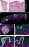
Figure 1
Figure 1
(A, B) Histological hallmarks of MF: intraepidermal vesicles (Pautrier’s microabscesses) and epidermotropism (atypical lymphocytes palisade along dermal-epidermal junction). (C) Slide setup with number of biopsies and type (NP=non-progression, PR=progression). (D) Photo of selected ROI/AOIs from one tissue sample. (E) ROI: dermal infiltrate, AOI: segmented into +/- CD4+ cells. (F) ROI: Pautrier’s microabscess. (G) ROI: Block of epidermis. (H) ROI: Basal layer.
Figure 2
Figure 2
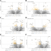
(A–E) A canonical visualization of the top five up- or downregulated genes for all five comparison models and known genes-of-interest (p<0.5) in progression vs. non-progression. None of the genes fell under the false detection rate (FDR) of 0.1. (F) All comparison models were combined in a total model showing the up- or downregulated genes enriched in progression vs. non-progression.
Figure 3
Figure 3
(A–E) Single-cell RNA sequencing (scRNA-seq) data from 76 skin biopsies (57 lesional, 7 non-lesional, 12 healthy) from 40 MF patients (stage I: six patients, stage II: 22 patients III: two patients, stage IV: eight patients, and two patients with unknown stage) and 12 healthy donors across six studies (–25) were integrated using scANVI and visualized using Uniform Manifold Approximation and Projection (UMAP) colored by (A) cell types, (B) Study or origin, (C) Biopsy site, (D) Expression of top T cell receptor (TCR)-clone marking malignant cells and (E) expression of KIR3DL2 known to be high in malignant cells in CTCL. (F) scRNA-seq expression of genes upregulated in patients with progression in the Pautrier’s microabscess model shown as percent positive cells and mean expression across identified cell types. (G–I). Transcript expression of the three selected genes (RUNX2, MCM7 and SHMT2) across each of the 76 Skin biopsies shown as transcripts per million (tpm) by sample. Bar height and colors indicate transcript contribution of each cell type population. (J) Mean malignant expression (as tpm) of RUNX2, MCM7 and SHMT2 divided into CTCL stage. Each dot represents mean malignant expression from one biopsy colored by lesion type. Bold horizontal line depicts median, box edges show 25th and 75th percentiles. The expression values indicated are gated on malignant T-cells only.

References

    1. Willemze R, Hodak E, Zinzani PL, Specht L, Ladetto M. Primary cutaneous lymphomas: ESMO Clinical Practice Guidelines for diagnosis, treatment and follow-up. Ann Oncol. (2013) 24:vi149–54. doi: 10.1093/annonc/mdt242 - DOI - PubMed
    1. Willemze R, Cerroni L, Kempf W, Berti E, Facchetti F, Swerdlow SH, et al. . The 2018 update of the WHO-EORTC classification for primary cutaneous lymphomas. Blood. (2019) 133:1703–14. doi: 10.1182/blood-2018-11-881268 - DOI - PMC - PubMed
    1. Kim YH, Liu HL, Mraz-Gernhard S, Varghese A, Hoppe RT. Long-term outcome of 525 patients with mycosis fungoides and sézary syndrome. Arch Dermatol. (2003) 139(7):857–866. doi: 10.1001/archderm.139.7.857 - DOI - PubMed
    1. Lindahl LM, Besenbacher S, Rittig AH, Celis P, Willerslev-Olsen A, Gjerdrum LMR, et al. . Prognostic miRNA classifier in early-stage mycosis fungoides: development and validation in a Danish nationwide study. Blood. (2018) 131:759–70. doi: 10.1182/blood-2017-06-788950 - DOI - PubMed
    1. Mourad A, Gniadecki R. Overall survival in mycosis fungoides: A systematic review and meta-analysis. J Invest Dermatol. (2020) 140:495–7. doi: 10.1016/j.jid.2019.07.712 - DOI - PubMed