Review
. 2025 Apr 1;40(4):632-640.
doi: 10.1093/ndt/gfae235.

The biology of water homeostasis

Affiliations
Review

The biology of water homeostasis

Mariavittoria D'Acierno et al. Nephrol Dial Transplant. .

Abstract

Water homeostasis is controlled by a brain-kidney axis that consists of central osmoreceptors, synthesis and secretion of arginine vasopressin (AVP) and AVP-responsive aquaporin-2 (AQP2) water channels in kidney collecting duct principal cells that facilitate water reabsorption. In addition to AVP, thirst represents a second line of defence to maintain water balance. Water balance disorders arise because of deficiency, resistance or inappropriate secretion of AVP or disturbances in thirst sensation (hypodipsia, polydipsia). People with water balance disorders are prone to develop hyponatraemia or hypernatraemia, which expose cells to osmotic stress and activate cell volume regulation mechanisms. This review covers several recent insights that have expanded our understanding of central osmoregulation, AQP2 regulation and cell volume regulation. This includes the role of with no lysine kinase 1 (WNK1) as a putative central osmolality sensor and, more generally, as an intracellular crowding sensor that coordinates the cell volume rescue response by activating sodium and potassium cotransporters. Furthermore, several new regulators of AQP2 have been identified, including AVP-dependent AQP2 regulation (yes-associated protein, nuclear factor of activated T-cells, microRNAs) and AVP-independent AQP2 regulation (epidermal growth factor receptor, fluconazole, prostaglandin E2). It is also becoming increasingly clear that long-term cell volume adaptation to chronic hypotonicity through release of organic osmolytes comes at the expense of compromised organ function. This potentially explains the complications of chronic hyponatraemia, including cognitive impairment, bone loss and vascular calcification. This review illustrates why these new insights derived from basic science are also relevant for developing new approaches to treat water balance disorders.

Keywords: aquaporin-2; cell volume regulation; hypernatraemia; hyponatraemia; vasopressin.


Conflict of interest statement

None declared.

Figures

Figure 1:
Figure 1:
Central osmoregulation. The brain regions involved in osmosensing, synthesis and secretion of AVP and thirst perception. Osmosensing neurons in the SFO and OVLT, both circumventricular organs, are capable of detecting an increase in plasma osmolality. The SFO interacts with the MnPO, which also receives signals from the oropharynx (fluid volume). SFO and OVLT neurons connect with the SON and PVN in the hypothalamus. The SON and PVN express magnocellular neurons that synthesize and secrete the hormones of the posterior pituitary, including AVP, apelin and oxytocin. Created with BioRender.com.
Figure 2:
Figure 2:
Water balance disorders. Water balance disorders arise as a consequence of disturbances in the secretion of or the kidneys’ response to AVP or disturbances in thirst perception. The left part of the figure shows the water balance disorders primary hypodipsia, AVP deficiency and AVP resistance, which are characterized by a risk of hypernatraemia. The right part of the figure shows the water balance disorders primary polydipsia and the syndrome of inappropriate antidiuresis, which can have a central or nephrogenic origin. These water balance disorders are characterized by a risk of hyponatraemia. AVP deficiency and AVP resistance are characterized by hypotonic urine, whereas the syndrome of inappropriate antidiuresis is characterized by hypertonic urine. Created with BioRender.com.
Figure 3:
Figure 3:
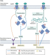
Modulators of AQP2 trafficking. The figure illustrates some of the key pathways involved in the regulation of AQP2 in collecting duct principal cells. In the canonical pathway, AVP activation of V2R results in greater cAMP levels, increased phosphorylation of AQP2 at Ser256 by PKA and translocation of AQP2 from intracellular vesicles to the apical membrane. Further phosphorylation of AQP2 at Ser269 increases AQP2 retention on the apical plasma membrane. cAMP-independent pathways are also important, and AQP2 can be phosphorylated by other kinases such as AMP-activated protein kinase (AMPK). AVP also modulates Rho-dependent remodelling of the actin cytoskeleton, which aids in AQP2 vesicle translocation. PGE2 alone acts through EP2 and EP4 receptors to enhance cAMP levels and AQP2 membrane accumulation. However, AVP stimulates expression of EP1 and/or EP3 receptors. Together EP1 and EP3 inhibit AQP2 translocation by stimulating RhoA and the formation of F-actin, alongside retrieval of AQP2 from the plasma membrane. In the absence of AVP, the ubiquitin E3 ligase CHIP promotes AQP2 ubiquitylation, internalization and lysosomal degradation. At a genetic level, the transcription cofactor YAP, through a complex consisting of GATA2, GATA3, NFATc1 and YAP/TEA domain transcription factor (TEAD) are critical for AQP2 transcription, which can be enhanced by AVP. The AQP2 gene can also be regulated by epigenetic factors because of alterations in principal cell miRNAs. Created with BioRender.com.
Figure 4:
Figure 4:
Vasopressin-independent modulators of aquaporin-2 trafficking. The figure illustrates some of the regulators of AQP2 plasma membrane targeting that are independent of the V2R and/or cAMP pathway. The EGFR inhibitor erlotinib enhanced AQP2 plasma membrane targeting and improved urine concentration in mice with lithium-induced AVP resistance. Metformin activates AMP-activated protein kinase (AMPK), leading to the phosphorylation of AQP2 at Ser256, increased AQP2 plasma membrane targeting and enhanced urine concentration in rats with tolvaptan-induced AVP resistance and in V2R knockout mice. Statins and fluconazole facilitate AQP2 plasma membrane targeting by inhibiting RhoA activity and actin polymerization. Created with BioRender.com.
Figure 5:
Figure 5:
Cell volume regulation. The effects of hypotonic and hypertonic stress on cell volume are depicted. Cells respond to hypotonic and hypertonic stress by activating cell volume regulation mechanisms to restore cell volume. WNK1 was identified as a sensor for a reduction in cell volume [42]. It is unclear if WNK1 can also sense cell swelling. In response to sensing a change in volume, cells activate channels and transporters to restore cell volume. Transport of water and organic osmolytes plays a role in both RVD and RVI. In RVD, potassium–chloride efflux also plays an important role, whereas in RVI, NKCC1 and chloride–bicarbonate transporters are activated. It is unclear if RVD and RVI are really orchestrated by a different set of transporters or if all transporters play a role in both processes. Created with BioRender.com.

References

    1. Bichet DG. Regulation of thirst and vasopressin release. Annu Rev Physiol 2019;81:359–73. 10.1146/annurev-physiol-020518-114556 - DOI - PubMed
    1. Augustine V, Gokce SK, Lee S et al. Hierarchical neural architecture underlying thirst regulation. Nature 2018;555:204–9. 10.1038/nature25488 - DOI - PMC - PubMed
    1. Matsuda T, Hiyama TY, Kobayashi K et al. Distinct CCK-positive SFO neurons are involved in persistent or transient suppression of water intake. Nat Commun 2020;11:5692. 10.1038/s41467-020-19191-0 - DOI - PMC - PubMed
    1. Kinsman BJ, Simmonds SS, Browning KN et al. Integration of hypernatremia and angiotensin II by the organum vasculosum of the lamina terminalis regulates thirst. J Neurosci 2020;40:2069–79. 10.1523/JNEUROSCI.2208-19.2020 - DOI - PMC - PubMed
    1. Zimmerman CA, Huey EL, Ahn JS et al. A gut-to-brain signal of fluid osmolarity controls thirst satiation. Nature 2019;568:98–102. 10.1038/s41586-019-1066-x - DOI - PMC - PubMed