. 2024 Oct 22;9(20):e179017.
doi: 10.1172/jci.insight.179017.

The STAT3/SETDB2 axis dictates NF-κB-mediated inflammation in macrophages during wound repair

Affiliations

The STAT3/SETDB2 axis dictates NF-κB-mediated inflammation in macrophages during wound repair

Kevin D Mangum et al. JCI Insight. .

Abstract

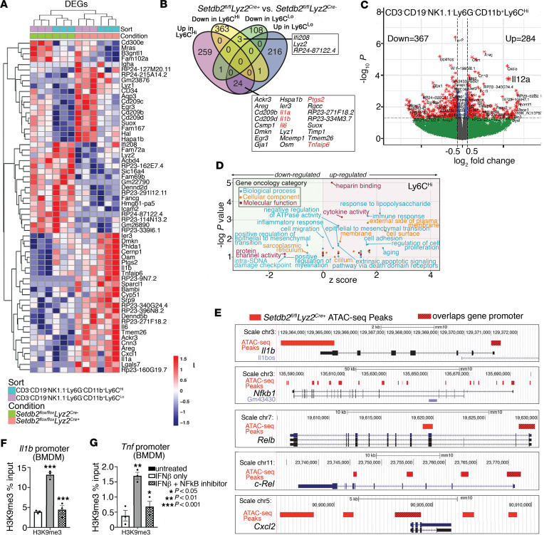
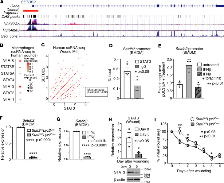
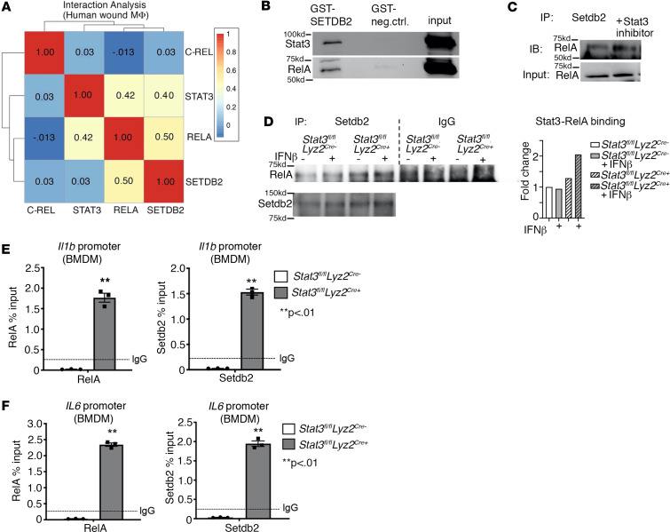
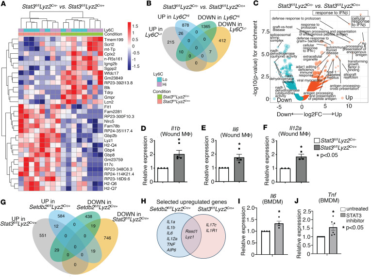
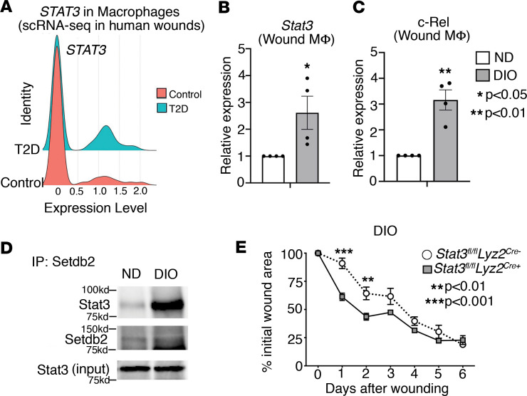
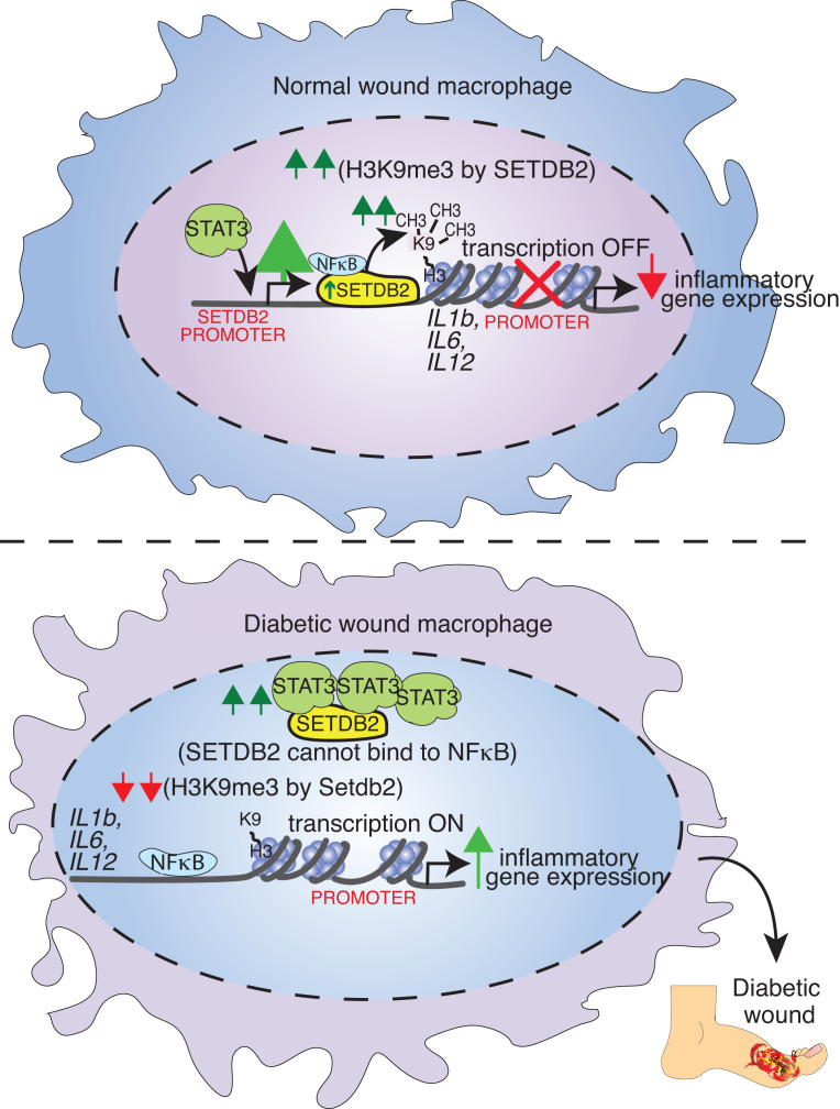
Macrophage transition from an inflammatory to reparative phenotype after tissue injury is controlled by epigenetic enzymes that regulate inflammatory gene expression. We have previously identified that the histone methyltransferase SETDB2 in macrophages drives tissue repair by repressing NF-κB-mediated inflammation. Complementary ATAC-Seq and RNA-Seq of wound macrophages isolated from mice deficient in SETDB2 in myeloid cells revealed that SETDB2 suppresses the inflammatory gene program by inhibiting chromatin accessibility at NF-κB-dependent gene promoters. We found that STAT3 was required for SETDB2 expression in macrophages, yet paradoxically, it also functioned as a binding partner of SETDB2 where it repressed SETDB2 activity by inhibiting its interaction with the NF-κB component, RELA, leading to increased RELA/NF-κB-mediated inflammatory gene expression. Furthermore, RNA-Seq in wound macrophages from STAT3-deficient mice corroborated this and revealed STAT3 and SETDB2 transcriptionally coregulate overlapping genes. Finally, in diabetic wound macrophages, STAT3 expression and STAT3/SETDB2 binding were increased. We have identified what we believe to be a novel STAT3/SETDB2 axis that modulates macrophage phenotype during tissue repair and may be an important therapeutic target for nonhealing diabetic wounds.

Keywords: Inflammation; Transcription.


Conflict of interest statement

Conflict of interest: The authors have declared that no conflict of interest exists.

Figures

Figure 1
Figure 1. SETDB2 is enriched at NF-κB–dependent gene promoters in murine wound macrophages.
Wound macrophages were isolated from Setdb2fl/fl Lyz2Cre mTmG murine wounds on day 5 after wounding, sorted into CD3CD19NK1.1Ly6GCD11b+Ly6Chi and CD3CD19NK1.1Ly6GCD11b+Ly6Clo populations, and then analyzed for gene expression by RNA-Seq and ATAC-Seq. (A) Heatmap of DEGs in wound macrophages isolated from Setdb2fl/fl Lyz2Cre mTmG murine wounds on day 5 after wounding. Wound macrophages were sorted into CD3CD19NK1.1Ly6GCD11b+Ly6Chi and CD3CD19NK1.1Ly6GCD11b+Ly6Clo populations and then analyzed for gene expression by RNA-Seq (12–15 mice per group, n = 3–5 independent experiments). (B) Venn diagram comparing upregulated and downregulated genes from RNA-Seq of wound macrophages (CD3CD19 NK1.1Ly6GCD11b+Ly6Chi and CD3CD19NK1.1Ly6GCD11b+Ly6Clo) isolated from Setdb2fl/fl Lyz2Cre mTmG mouse wounds on day 5 after wounding. (C) Volcano plot of transcriptomic profiles from DEGs in CD3CD19NK1.1Ly6GCD11b+Ly6Chi wound macrophages from Setdb2fl/fl Lyz2Cre mTmG mice. (D) GO analysis of DEGs in CD3CD19NK1.1Ly6GCD11b+Ly6Chi wound macrophages from Setdb2fl/fl Lyz2Cre mTmG mice. (E) Inflammatory gene promoter regions differentially regulated by SETDB2 as determined by ATAC-Seq of wound macrophages (CD3CD19NK1.1Ly6GCD11b+Ly6C+ GFP+) isolated on day 5 from Setdb2fl/fl Lyz2Cre mTmG mice. Red bars indicate ATAC-Seq peaks relative to Cre-negative controls. Hashed red bars designate peaks that overlap promoter regions of listed genes. (F and G) H3K9me3 ChIP qPCR at the Il1b and Tnf promoters in murine BMDMs unstimulated or treated with IFN-β (10 U/mL; 8.5 ng/mL) or IFN-β and NF-κB inhibitor, BAY 11-7082 (10 μM). Data are representative of n = 3–5 independent experiments, with 12–15 mice per group per experiment. *P < 0.05, **P < 0.01, ***P < 0.001. Data are presented as the mean ± SEM. Two-tailed Student’s t test was used for comparison of 2 groups. For comparison among multiple groups, 2-way ANOVA followed by Newman-Keuls post hoc test was used.
Figure 2
Figure 2. STAT3 is dynamic during wound repair and regulates SETDB2 in human and murine wound macrophages.
(A) Gene structure of the human SETDB2 gene showing cloned fragment aligned with multiple active transcriptional features including DHS peaks, H3K27Ac, H3K4me3, and sequence conservation. (B) Dot plot of different STAT members expression from scRNA-Seq data of human wounds (n = 10 patients). (C) Scatterplot of SETDB2 and STAT3 expression in macrophages obtained from scRNA-Seq in human wounds with respective Pearson correlation analysis (n = 10 patients). (D) ChIP-qPCR for Stat3 binding at the mouse Setdb2 promoter compared with IgG negative control. (E) Luciferase activity of the 3 kb human SETDB2 promoter cloned into pGL3 and then transfected in BMDMs untreated or treated with IFN-β (10 U/mL; 8.5 ng/mL), or IFN-β plus tofacitinib (100 μM) for 4 hours. (F) qPCR analysis of Setdb2 expression in wound macrophages (CD3CD19NK1.1Ly6GCD11b+) isolated from Stat3fl/fl Lyz2Cre mice on day 5 after wounding compared with Cre littermate control (n = 6 mice per group). (G) Setdb2 expression in BMDMs treated with IFN-β or IFN-β plus tofacitinib (n = 6–8 mice per group). (H) Representative Western blot and densitometry of murine whole wounds showing decreased levels of STAT3 at day 5 compared with day 0 after wounding (n = 3–4 mice at each time point). (I) Wound curve analysis in Stat3fl/fl Lyz2Cre mice compared with Cre littermate controls all fed a normal diet (n = 8–12 mice per group in each experiment). All data are representative of n = 3–5 independent experiments. *P < 0.05, **P < 0.01, ****P < 0.0001. Data are presented as the mean ± SEM. Two-tailed Student’s t test was used for comparison of 2 groups. For comparison among multiple groups, 2-way ANOVA followed by Newman-Keuls post hoc test was used.
Figure 3
Figure 3. STAT3 inhibits the physical interaction between SETDB2 and NF-κB in wound macrophages.
(A) Heatmap of Pearson gene correlation analysis results for SETDB2, STAT3, C-REL, and RELA in macrophages obtained from scRNA-Seq in human wounds (n = 10 patients). (B) Western blot showing Setdb2-interacting proteins purified after incubating GST-SETDB2 with BMDM lysate. (C) Western blot of RelA coimmunoprecipitated with Setdb2 from BMDMs treated with Stat3 inhibitor compared with untreated BMDMs. (D) Setdb2 immunoprecipitate reactions from BMDMs isolated from Stat3fl/fl Lyz2Cre+ mice and Cre-negative littermate controls that were untreated or treated with IFN-β (10 U/mL; 8.5 ng/mL) for 4 hours were analyzed via Western blot and probed for RelA and Setdb2 (n = 4–6 mice per group). (E and F) ChIP-qPCR on BMDMs isolated from Stat3fl/fl Lyz2Cre+ versus Cre-negative controls for NF-κB (RelA) and Setdb2 at the Il1b and Il6 promoters (n = 4–6 mice per group). All data are representative of n = 3–5 independent experiments. **P < 0.01. Data are presented as the mean ± SEM. Two-tailed Student’s t test was used for comparison of 2 groups. For comparison among multiple groups, 2-way ANOVA followed by Newman-Keuls post hoc test was used.
Figure 4
Figure 4. STAT3 and SETDB2 coregulate inflammatory gene expression in wound macrophages.
(A) Heatmap of DEGs in day 5 wound macrophages (CD3CD19NK1.1Ly6GCD11b+) from Stat3fl/fl Lyz2Cre mice and Cre littermate controls (n = 6 mice per group). (B) Diagram comparing overlapping genes among upregulated and downregulated genes from RNA-Seq of Stat3fl/fl Lyz2Cre wound macrophages (CD3CD19NK1.1Ly6GCD11b+). (C) Volcano plot of GO analysis for the enriched biological processes in decreased and increased DEGs from Stat3fl/fl Lyz2Cre wound macrophages. (DF) qPCR of Il1b (D), Il6 (E), and Il12a (F) in of Stat3fl/fl Lyz2Cre BMDMs treated with IFN-β (10 U/mL; 8.5 ng/mL) for 6 hours (n = 5 mice per group). (G) Diagram demonstrating overlapping genes among upregulated and downregulated DEGs from RNA-Seq of Setdb2fl/fl Lyz2Cre and Stat3fl/fl Lyz2Cre CD3CD19NK1.1Ly6GCD11b+ macrophages isolated from day 5 wounds. (H) Venn diagram of overlapping and distinct upregulated inflammatory genes in the CD3CD19NK1.1Ly6GCD11b+Ly6Chi population from wounds isolated on day 5 from Setdb2fl/fl Lyz2Cre and Stat3fl/fl Lyz2Cre mice. (I and J) mRNA expression of Il6 and Tnf in BMDMs treated with a STAT3 inhibitor compared with untreated (n = 5 mice per group in each experiment). All data are representative of n = 3–5 independent experiments. *P < 0.05. Data are presented as the mean ± SEM. Two-tailed Student’s t test was used for comparison of 2 groups. For comparison among multiple groups, 2-way ANOVA followed by Newman-Keuls post hoc test was used.
Figure 5
Figure 5. STAT3 is increased in diabetic wound macrophages during the transition to the reparative phenotype.
(A) scRNA-Seq analysis of STAT3, C-REL, and RELA in macrophages from human wounds isolated from nondiabetic and diabetic patients (n = 10 patients). (B and C) qPCR of Stat3 and c-Rel expression on day 0 and 5 after wounding in ND and DIO murine wound macrophages (CD3CD19NK1.1Ly6GCD11b+) treated with IFN-β (10 U/mL; 8.5 ng/mL) for 6 hours ex vivo (n = 12–16 mice per group). (D) Coimmunoprecipitation of endogenous Setdb2 and Stat3 from ND and DIO whole wounds isolated day 5 after wounding (n = 4 mice per group). (E) Wound curve analysis in Stat3fl/fl Lyz2Cre DIO mice compared with Cre littermate DIO controls (n = 8–12 mice per group). All data are representative of n = 3–5 independent experiments. *P < 0.05, **P < 0.01, ***P < 0.001. Data are presented as the mean ± SEM. Two-tailed Student’s t test was used for comparison of 2 groups. For comparison among multiple groups, 2-way ANOVA followed by Newman-Keuls post hoc test was used.
Figure 6
Figure 6. Schematic of the STAT3/NF-κB/SETDB2 axis in normal and diabetic wound macrophages.

References

    1. Medzhitov R. Origin and physiological roles of inflammation. Nature. 2008;454(7203):428–435. doi: 10.1038/nature07201. - DOI - PubMed
    1. Wynn TA, Vannella KM. Macrophages in tissue repair, regeneration, and fibrosis. Immunity. 2016;44(3):450–462. doi: 10.1016/j.immuni.2016.02.015. - DOI - PMC - PubMed
    1. Wolf J, et al. Interleukin-6 and its receptors: a highly regulated and dynamic system. Cytokine. 2014;70(1):11–20. doi: 10.1016/j.cyto.2014.05.024. - DOI - PubMed
    1. Kittan NA, et al. Cytokine induced phenotypic and epigenetic signatures are key to establishing specific macrophage phenotypes. PLoS One. 2013;8(10):e78045. doi: 10.1371/journal.pone.0078045. - DOI - PMC - PubMed
    1. Alexandraki KI, et al. Cytokine secretion in long-standing diabetes mellitus type 1 and 2: associations with low-grade systemic inflammation. J Clin Immunol. 2008;28(4):314–321. doi: 10.1007/s10875-007-9164-1. - DOI - PubMed

MeSH terms