Serial Optical Coherence Tomography Assessment of Coronary Atherosclerosis and Long-Term Clinical Outcomes
- PMID: 39435729
- PMCID: PMC11935709
- DOI: 10.1161/JAHA.123.034458
Serial Optical Coherence Tomography Assessment of Coronary Atherosclerosis and Long-Term Clinical Outcomes
Abstract
Background: The impact of high-risk coronary artery plaques identified using optical coherence tomography on late luminal narrowing and clinical events remains poorly understood.
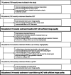
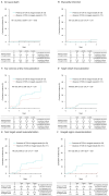
Methods and results: This multicenter prospective study included 176 patients who underwent percutaneous coronary intervention and serial optical coherence tomography at baseline and 1-year follow-up to investigate nontarget regions with angiographically intermediate stenosis. At 1 year after percutaneous coronary intervention, the coronary artery lumen area decreased significantly from 6.06 (95% CI, 5.60-6.53) mm2 to 5.88 (95% CI, 5.41-6.35) mm2 (difference, -0.18; 95% CI, -0.22 to -0.14 mm2; P<0.001), particularly in thin-cap fibroatheromas, thick-cap fibroatheromas, mixed plaques, and fibrous plaques. The prevalence of fibroatheroma decreased from 38% to 36% (P<0.001), whereas calcified plaque increased from 31% to 34% (P<0.001), accompanied by a significant increase in calcium thickness and angle. Diabetes and current smoking habits were independently associated with increasing calcium prevalence. Patients with thin-cap fibroatheroma had a significantly higher 3-year risk of ischemia-driven nontarget vessel revascularization (hazard ratio, 2.42 [95% CI, 1.03-5.71]; P=0.04), primarily due to revascularization in the imaged region. No significant association was observed between coronary artery calcium prevalence and clinical outcomes within 3 years.
Conclusions: The coronary artery lumen area significantly decreased over a 1-year interval, particularly in thin-cap fibroatheromas, thick-cap fibroatheromas, mixed plaques, and fibrous plaques. Although thin-cap fibroatheroma prevalence was associated with higher risk of ischemia-driven nontarget vessel revascularization, no significant association was noted between coronary artery calcium prevalence and clinical outcomes within 3 years. The interaction between calcium progression and long-term clinical events necessitates further investigation.
Registration: URL: https://www.umin.ac.jp/ctr/; Unique Identifier: UMIN000031937.
Keywords: atherosclerotic; coronary artery disease; optical coherence tomography.
Figures


Comment in
-
Following the Dynamic Changes of Coronary Atherosclerosis: An Uphill Battle.J Am Heart Assoc. 2024 Nov 5;13(21):e037395. doi: 10.1161/JAHA.124.037395. Epub 2024 Oct 22. J Am Heart Assoc. 2024. PMID: 39435716 Free PMC article. No abstract available.
References
-
- Yamaji K, Shiomi H, Morimoto T, Toyota T, Ono K, Furukawa Y, Nakagawa Y, Kadota K, Ando K, Shirai S, et al. Influence of sex on long‐term outcomes after implantation of bare‐metal stent: a multicenter report from the coronary revascularization demonstrating outcome study‐Kyoto (CREDO‐Kyoto) registry Cohort‐1. Circulation. 2015;132:2323–2333. doi: 10.1161/CIRCULATIONAHA.115.017168 - DOI - PubMed
-
- Maehara A, Cristea E, Mintz GS, Lansky AJ, Dressler O, Biro S, Templin B, Virmani R, de Bruyne B, Serruys PW, et al. Definitions and methodology for the grayscale and radiofrequency intravascular ultrasound and coronary angiographic analyses. JACC Cardiovasc Imaging. 2012;5:S1–S9. doi: 10.1016/j.jcmg.2011.11.019 - DOI - PubMed
Publication types
MeSH terms
LinkOut - more resources
Full Text Sources
Medical
Miscellaneous

