. 2024 Nov 4;156(11):e202313530.
doi: 10.1085/jgp.202313530. Epub 2024 Oct 22.

Light induces a rapid increase in cAMP and activates PKA in rod outer segments of the frog retina

Affiliations

Light induces a rapid increase in cAMP and activates PKA in rod outer segments of the frog retina

Olga Chernyshkova et al. J Gen Physiol. .

Abstract

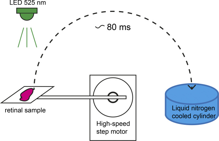
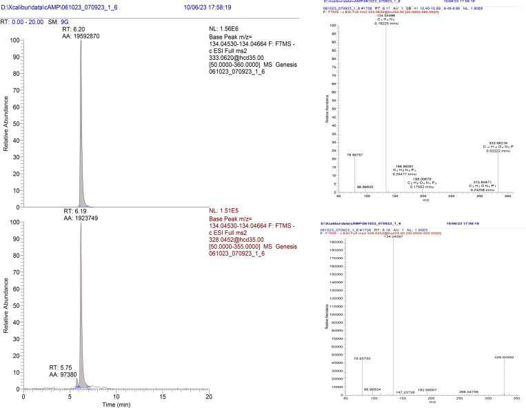
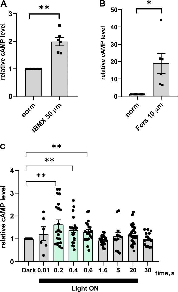
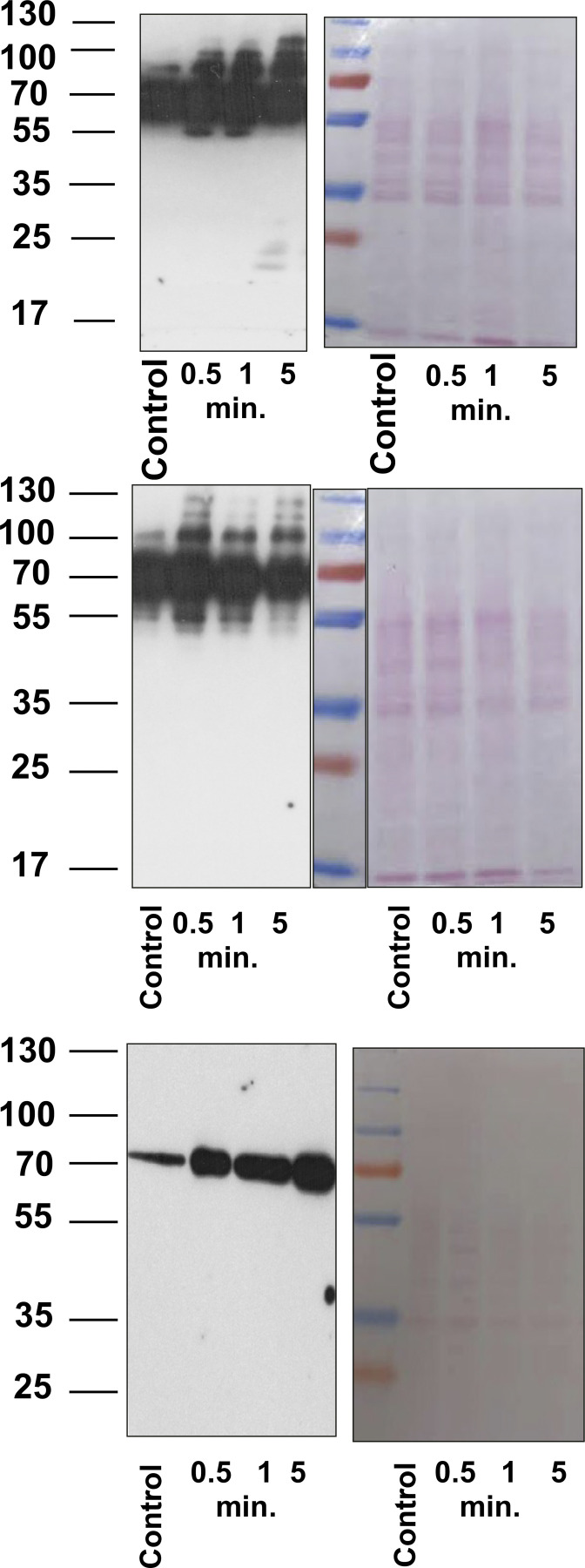
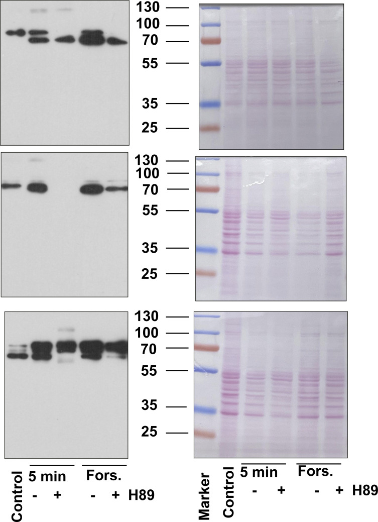
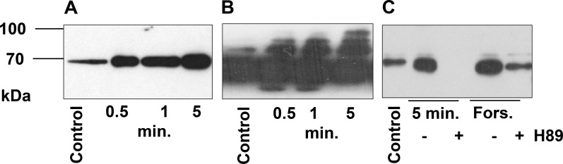
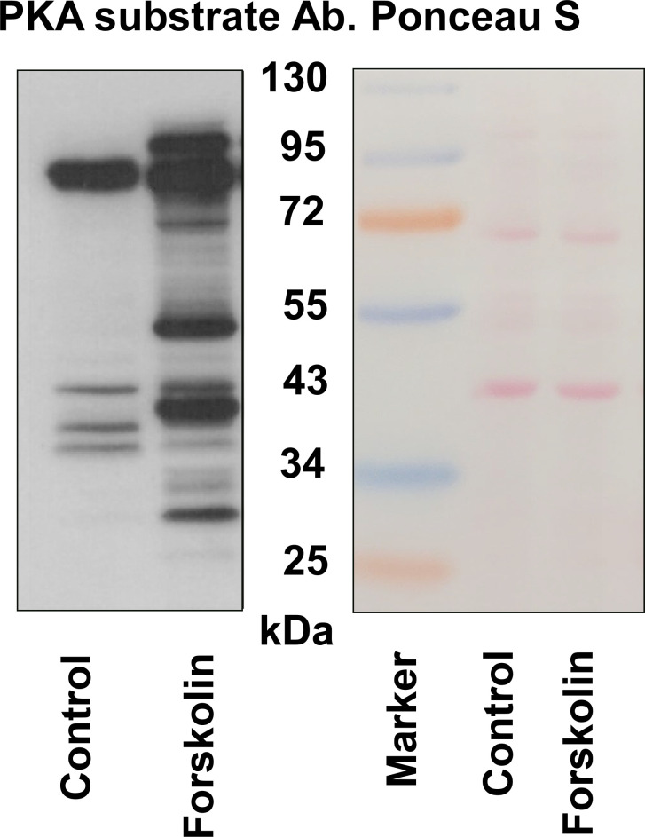
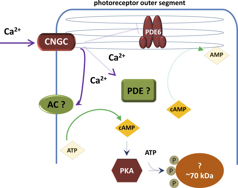
The phototransduction cascade enables the photoreceptor to detect light over a wide range of intensities without saturation. The main second messenger of the cascade is cGMP and the primary regulatory mechanism is calcium feedback. However, some experimental data suggest that cAMP may also play a role in regulating the phototransduction cascade, but this would require changes in cAMP on a time scale of seconds. Currently, there is a lack of data on the dynamics of changes in intracellular cAMP levels on this timescale. This is largely due to the specificity of the sensory modality of photoreceptors, which makes it practically impossible to use conventional experimental approaches based on fluorescence methods. In this study, we employed the method of rapid cryofixation of retinal samples after light stimulation and subsequent isolation of outer segment preparations. The study employed highly sensitive metabolomics approaches to measure levels of cAMP. Additionally, PKA activity was measured in the samples using a western blot. The results indicate that when exposed to near-saturating but still moderate light, cAMP levels increase transiently within the first second and then return to pre-stimulus levels. The increase in cAMP activates PKA, resulting in the phosphorylation of PKA-specific substrates in frog retinal outer segments.


Conflict of interest statement

Disclosures: D. Meshalkina reported grants from the Russian Ministry of Science and Education project 075-15-2022-296 World-class research center Pavlov Center “Integrative Physiology to Medicine, High-Tech Healthcare and Technologies of Stress Resistance” outside the submitted work. No other disclosures were reported.

Figures

Figure 1.
Figure 1.
Schematic diagram of the unit for cryofixation of retinal preparation. After light stimulation, a stepper motor moves the retina at 80 ms and presses it against a polished copper cylinder in liquid nitrogen.
Figure S1.
Figure S1.
Example of chromatograms and mass spectra of cAMP (lower) and internal standard isotope-labeled cAMP (upper) in a frog ROS sample.
Figure 2.
Figure 2.
cAMP content in frog retina rod outer segments. (A and B) cAMP level in ROS after incubation for 5 min in 50 µM IBMX Ringer solution (A) or incubation for 20 min in 10 µM Forskolin Ringer solution (B). Data are relative to cAMP content in normal Ringer’s solution in the dark. (C) Concentration of cAMP in ROS after exposure to light of different durations (from 0.01 to 20 s) and after extinction of the stimulus of 20 s duration. Intensity 4.6 × 10⁴ quanta/(s·µm2). The black horizontal bar on the graph marks the beginning and end of light stimulation. All values are shown as relative to the measurement of the dark-adapted sample on the same eye or on the same frog. Error bars indicate SEM and stars indicate significant difference (one-way ANOVA with Dunnett’s multiple comparisons test).
Figure S2.
Figure S2.
Full blots and Ponceau S stained membrane of three independent experiments. Source data are available for this figure: SourceDataFS2.
Figure S3.
Figure S3.
Full blots and Ponceau S stained membranes of three independent experiments. Source data are available for this figure: SourceDataFS3.
Figure 3.
Figure 3.
Light stimulation activates PKA in frog outer segments. (A and B) Frozen sections of outer segments in the dark (control) and after 0.5, 1, and 5 min of light stimulation were analyzed by western blotting with PKA substrate antibody (A and B). (C) Retina were preincubated with PKA inhibitor H89 (50 µM, 10 min) and then stimulated with forskolin (forskolin samples) or light-stimulated for 5 min. 10 µg of protein were loaded in each lane. The signal was visualized by ECL detection. A and B are the same blots exposed during 1 s (A) and 10 s (B). Light intensity is the same as in Fig. 2 C. Equal loading was controlled by Ponceau S staining of the membranes (Fig. S2 and Fig. S3). Source data are available for this figure: SourceData F3.
Figure S4.
Figure S4.
Isolated human platelets (3 × 10⁷/lane) without stimulation (control) and stimulated by forskolin (5 µM, 2 min) were prepared for western blot analysis with PKA substrate antibody. Membrane was stained by Ponceau S. Source data are available for this figure: SourceDataFS4.
Figure 4.
Figure 4.
Hypothetical scheme of cAMP turnover in the outer segment of a photoreceptor cell. CNGC – cyclic nucleotide (cGMP) gated channel, AC – adenylate cyclase, PDE – phosphodiesterase, PKA – cAMP-dependent protein kinase. Approximately, 70 kDa – unidentified protein that undergoes phosphorylation upon PKA activation.

References

    1. Astakhova, L.A., Kapitskii S.V., Govardovskii V.I., and Firsov M.L.. 2014. Cyclic AMP as a regulator of the phototransduction cascade. Neurosci. Behav. Physiol. 44:664–671. 10.1007/s11055-014-9967-5 - DOI
    1. Astakhova, L.A., Samoiliuk E.V., Govardovskii V.I., and Firsov M.L.. 2012. cAMP controls rod photoreceptor sensitivity via multiple targets in the phototransduction cascade. J. Gen. Physiol. 140:421–433. 10.1085/jgp.201210811 - DOI - PMC - PubMed
    1. Baylor, D.A., Lamb T.D., and Yau K.W.. 1979. Responses of retinal rods to single photons. J. Physiol. 288:613–634. 10.1113/jphysiol.1979.sp012716 - DOI - PMC - PubMed
    1. Beck, F., Geiger J., Gambaryan S., Veit J., Vaudel M., Nollau P., Kohlbacher O., Martens L., Walter U., Sickmann A., and Zahedi R.P.. 2014. Time-resolved characterization of cAMP/PKA-dependent signaling reveals that platelet inhibition is a concerted process involving multiple signaling pathways. Blood. 123:e1–e10. 10.1182/blood-2013-07-512384 - DOI - PubMed
    1. Benovic, J.L. 2021. Historical perspective of the G protein-coupled receptor kinase family. Cells. 10:555. 10.3390/cells10030555 - DOI - PMC - PubMed

Substances

LinkOut - more resources