. 2024 Oct 29;121(44):e2321305121.
doi: 10.1073/pnas.2321305121. Epub 2024 Oct 22.

eIF4F controls ERK MAPK signaling in melanomas with BRAF and NRAS mutations

Affiliations

eIF4F controls ERK MAPK signaling in melanomas with BRAF and NRAS mutations

Barbora Valcikova et al. Proc Natl Acad Sci U S A. .

Abstract

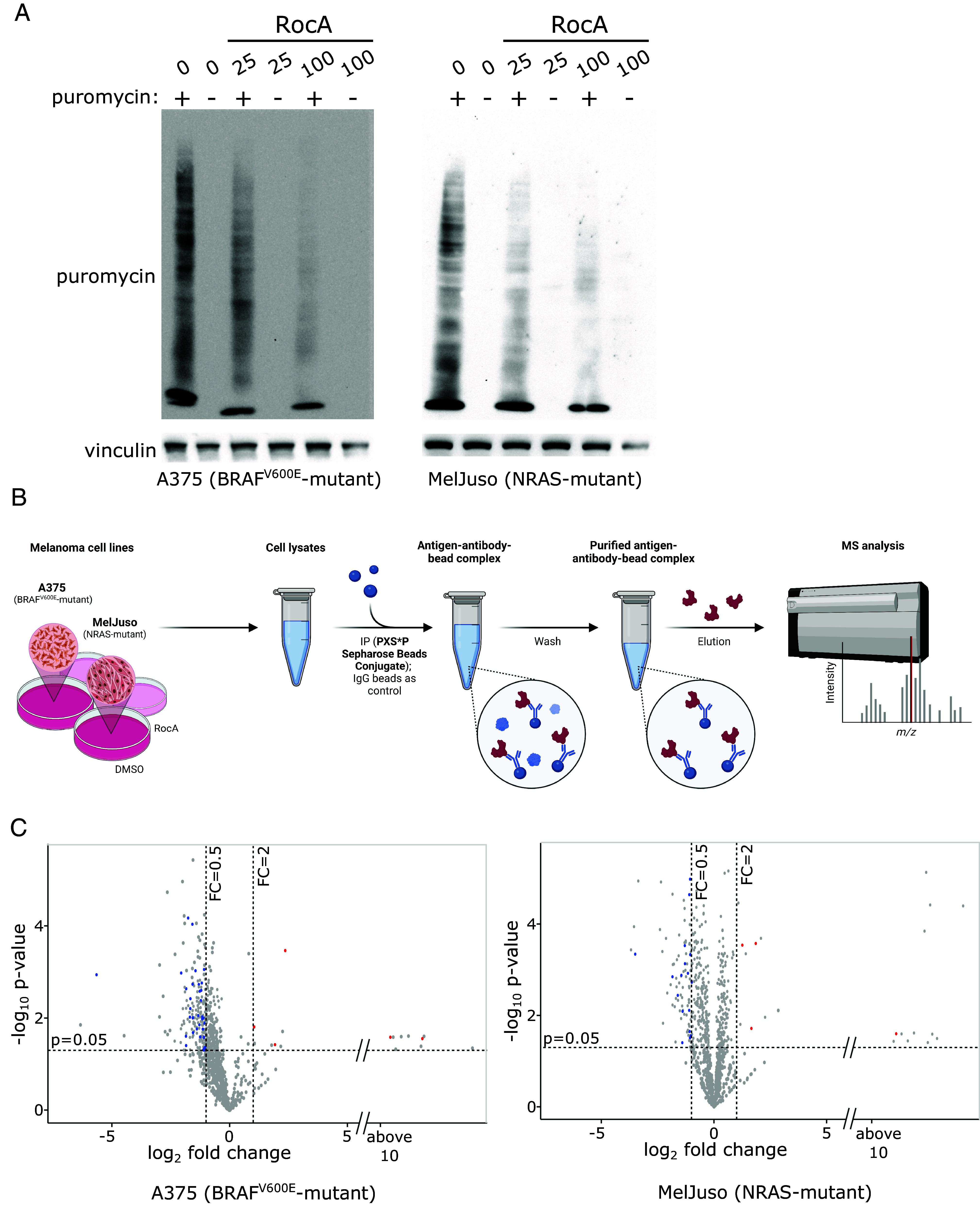
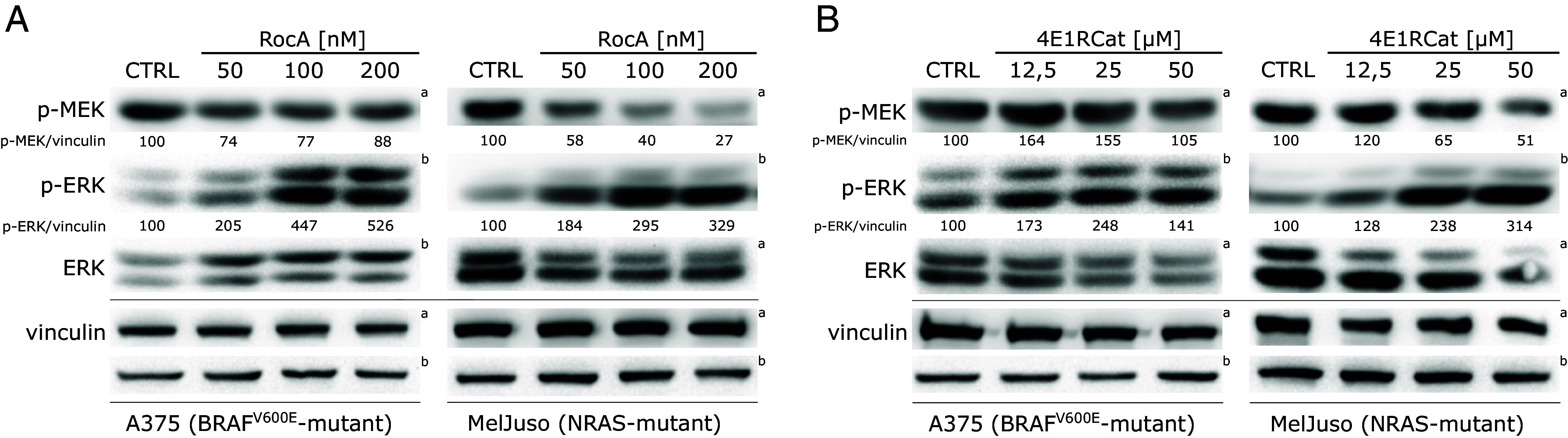
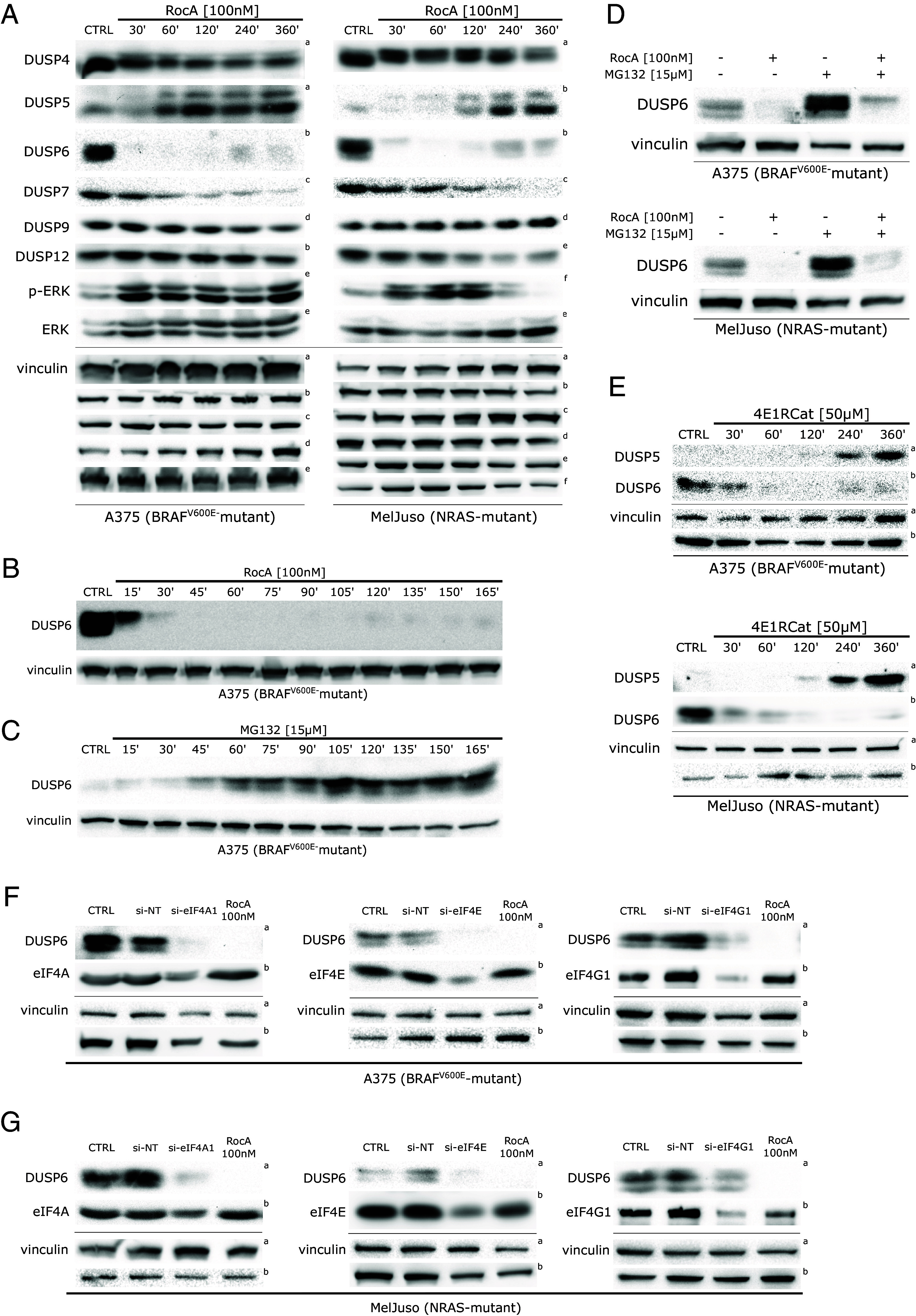
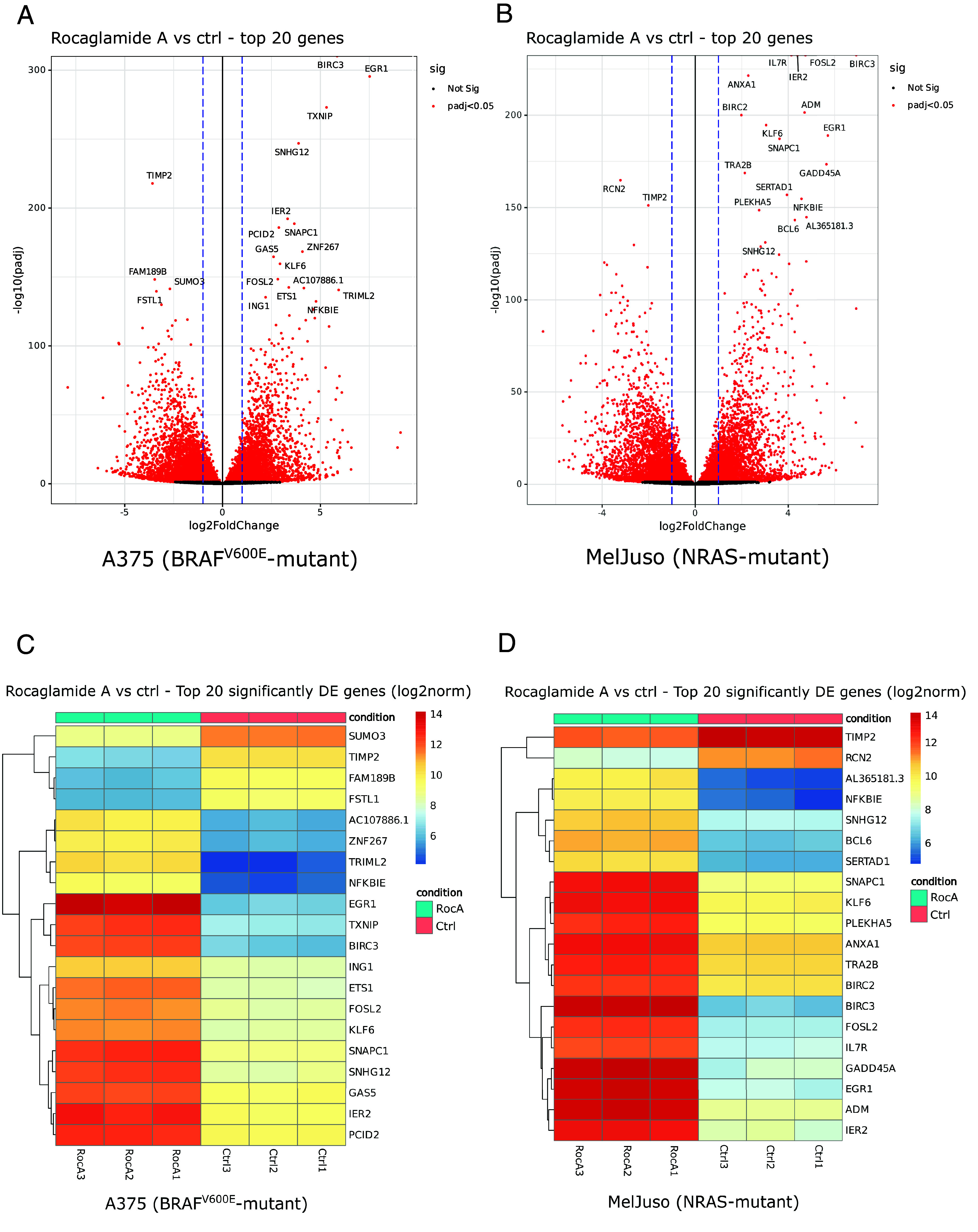
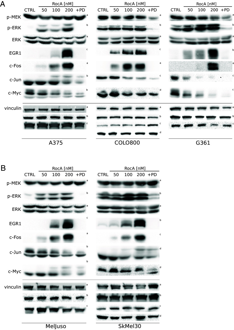
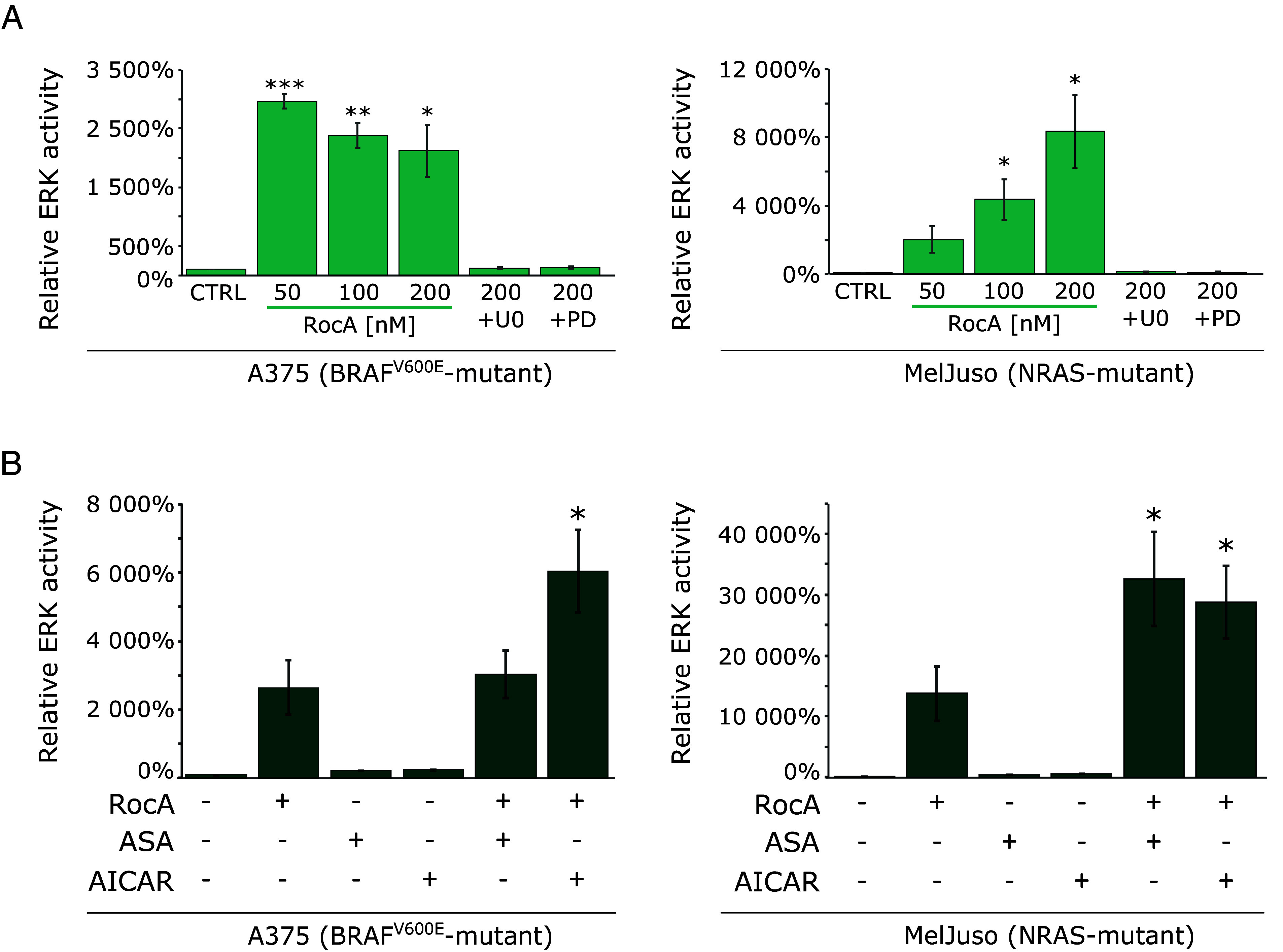
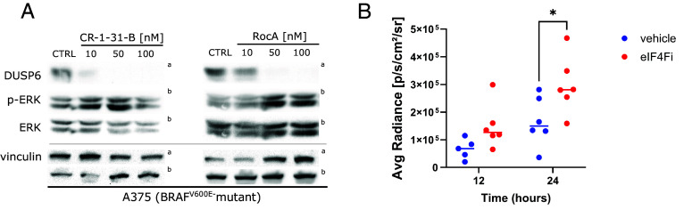
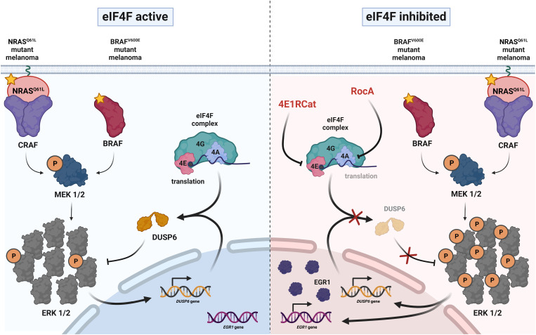
The eIF4F translation initiation complex plays a critical role in melanoma resistance to clinical BRAF and MEK inhibitors. In this study, we uncover a function of eIF4F in the negative regulation of the rat sarcoma (RAS)/rapidly accelerated fibrosarcoma (RAF)/mitogen-activated protein kinase kinase (MEK)/extracellular signal-regulated kinase (ERK) mitogen-activated protein kinase (MAPK) signaling pathway. We demonstrate that eIF4F is essential for controlling ERK signaling intensity in treatment-naïve melanoma cells harboring BRAF or NRAS mutations. Specifically, the dual-specificity phosphatase DUSP6/MKP3, which acts as a negative feedback regulator of ERK activity, requires continuous production in an eIF4F-dependent manner to limit excessive ERK signaling driven by oncogenic RAF/RAS mutations. Treatment with small-molecule eIF4F inhibitors disrupts the negative feedback control of MAPK signaling, leading to ERK hyperactivation and EGR1 overexpression in melanoma cells in vitro and in vivo. Furthermore, our quantitative analyses reveal a high spare signaling capacity in the ERK pathway, suggesting that eIF4F-dependent feedback keeps the majority of ERK molecules inactive under normal conditions. Overall, our findings highlight the crucial role of eIF4F in regulating ERK signaling flux and suggest that pharmacological eIF4F inhibitors can disrupt the negative feedback control of MAPK activity in melanomas with BRAF and NRAS activating mutations.

Keywords: DUSP6; ERK; MAP kinase; eIF4F; melanoma.


Conflict of interest statement

Competing interests statement:The authors declare no competing interest.

Figures

Fig. 1.
Fig. 1.
In melanoma cells, a proportion of MAPK targets are synthesized in an eIF4F-dependent manner. (A) The effect of eIF4F inhibition on the rate of protein synthesis in melanoma cells was analyzed using the puromycylation assay. A375 (BRAFV600E) and MelJuso (NRAS-mutant) cells were treated with the indicated concentrations of Rocaglamide A (RocA) for 4 h. Next, newly synthesized peptide chains were covalently labeled by adding puromycin (90 µM final concentration) to the cell cultures for 15 min, followed by cell lysis, SDS-PAGE, and Western blotting. Vinculin served as a loading control. The controls were treated with an equivalent volume of the vehicle (DMSO). (B) Schematic representation of the identification of MAPK targets responding to eIF4F inhibition. A375 and MelJuso cells were treated with the eIF4F inhibitor Rocaglamide A (RocA, 100 nM) for 20 h. Phospho-MAPK/CDK Substrate Motif (PXS*P and S*PXK/R) Kit was used for the immunoprecipitation of cell lysates, followed by the LC-MS/MS analysis to identify potential MAPK targets affected by eIF4F inhibition. Created with BioRender.com. (C) A proportion of MAPK targets is downregulated in response to eIF4F inhibition in both melanoma subtypes. Volcano plots depict changes in protein levels in cells treated with Rocaglamide A (RocA, 100 nM) for 20 h. Previously identified ERK targets among the downregulated proteins are depicted in blue and characterized by log2 Fold Change < −0.5 and P-value < 0.05. Red dots represent up-regulated previously identified ERK targets (http://sys-bio.net/erk_targets/targets_all.html), characterized by log2 Fold Change > 2 and P-value < 0.05.
Fig. 2.
Fig. 2.
eIF4F inhibition promotes ERK activation in melanoma cells. Western blot analysis of A375 and MelJuso cells after 20 h treatment with increasing concentrations of small-molecule eIF4F inhibitors. (A) eIF4A inhibitor Rocaglamide A (RocA) or (B) eIF4E-eIF4G disruptor, 4E1RCat. The activity of MEK and ERK is represented by the ratio of phospho-MEK and phospho-ERK (p-MEK, p-ERK) to vinculin levels. Vinculin served as a loading control. The control samples (CTRL) were treated with an equivalent volume of the vehicle (DMSO). The upper index letters refer to the corresponding loading control detected on the same membrane.
Fig. 3.
Fig. 3.
ERK activity in melanoma is limited by eIF4F-dependent expression of DUSP6/MKP3. (A) eIF4A inhibition induces dynamic changes in the levels of the DUSP/MKP family members, which function as critical negative regulators of the ERK pathway. Western blot analysis of A375 and MelJuso cells treated with RocA for indicated time periods. The upper index letters refer to the corresponding loading control detected on the same membrane. (B) Western blot analysis of A375 cells treated with Rocaglamide A (RocA) in shorter time periods shows the rapid degradation of DUSP6/MKP3 in response to eIF4F inhibition. (C) Western blot analysis of A375 cells treated with 15 µM MG132 showing the accumulation of DUSP6/MKP3 in response to proteasome inhibition. (D) The addition of a proteasome inhibitor led to a partial recovery of DUSP6/MKP3 levels caused by eIF4F inhibition alone, as shown in Western blot analysis of A375 and MelJuso cells treated with Rocaglamide A and/or MG132 for 16 h. (E) A structurally unrelated small-molecule eIF4Fi also promoted dynamic changes in the levels of DUSP6 and DUSP5. Western blot analysis was performed on lysates of A375 and MelJuso cells treated with 50 µM 4E1RCat for indicated periods. The upper index letters refer to the corresponding loading control detected on the same membrane. Vinculin served as a loading control. The control samples (CTRL) were treated with an equivalent volume of the vehicle (DMSO). (F and G) Knockdown of individual eIF4F subunits recapitulates the impact of small-molecule eIF4F inhibitors on DUSP6 expression in melanoma cells. Western blot analysis of A375 (F) and MelJuso (G) cells transiently transfected with siRNAs specific for eIF4A1, eIF4E, and eIF4G1 for 48 h. Non-targeting siRNAs (si-NT) were transfected in parallel as negative controls. 24 h treatment with RocA served as a positive control, while the control samples (CTRL) were treated with an equivalent volume of the vehicle (DMSO). The upper index letters refer to the corresponding loading control detected on the same membrane.
Fig. 4.
Fig. 4.
Changes in melanoma cell transcriptome in response to eIF4F inhibition. We used next-generation RNA sequencing to analyze transcriptome changes in response to 20 h RocA treatments. Volcano plots show overall changes in gene expression in A375 (A) and MelJuso (B) melanoma cells in response to eIF4Fi. Heat maps present the response to eIF4Fi of the top 20 differentially expressed genes in the individual independent biological replicates [A375 (C), MelJuso (D)].
Fig. 5.
Fig. 5.
In a panel of melanoma cell lines, eIF4F inhibition promotes the hyperactivation of the ERK pathway, along with ERK-driven upregulation of EGR1 and c-Fos transcription factors. ERK activity and the levels of selected ERK downstream targets were analyzed by Western blot in (A) BRAFV600E-mutant (A375, COLO800, G361) and (B) NRAS-mutant (MelJuso, SKMel30) melanoma cells in response to a 20 h treatment with Rocaglamide A (RocA) at indicated concentrations. A combination of RocA and PD184352, a small-molecule MEK inhibitor (PD, 0.5 µM), confirmed that EGR1 and c-Fos upregulation is caused selectively by ERK hyperactivation. Vinculin served as a loading control. The control samples (CTRL) were treated with an equivalent volume of the vehicle (DMSO). The upper index refers to the corresponding loading control detected on the same membrane.
Fig. 6.
Fig. 6.
ERK pathway hyperactivation caused by eIF4F inhibition is amplified under metabolic stress conditions. (A) Quantitative analysis of the relative ERK activity in A375 and MelJuso cells increased in response to eIF4F inhibition. Cells, stably transfected with the reporter construct pKROX24(MapErk)Luc, expressing firefly luciferase under the control of an EGR1 gene-derived promoter, were treated with Rocaglamide A alone or in combination with MEK inhibitors, U0126 (U0, 10 µM) or PD184352 (PD, 0.5 µM) for 24 h. (B) eIF4F inhibition and AMPK activation synergize in increasing the signaling capacity of the ERK pathway. A375 and MelJuso cells stably transfected with pKROX24(MapErk)Luc were treated with Rocaglamide A (200 nM) alone or in combination with AMPK activators Acetylsalicylic Acid (ASA, 5 mM) and 5-Aminoimidazole-4-carboxamide 1-β-D-ribofuranoside (AICAR, 2 mM) for 24 h. The control samples (CTRL) were treated with an equivalent volume of the vehicle (DMSO). Results were obtained from three independent repetitions and normalized to protein concentration. The data are presented by an average of ERK activity relative to the control samples ± SE. *P < 0.05.
Fig. 7.
Fig. 7.
eIF4F inhibition promotes ERK hyperactivation in melanoma tumors in vivo. (A) The synthetic rocaglate CR-1-31-B downregulates DUSP6 and promotes ERK hyperactivation in A375 melanoma in vitro. Cells were treated with the indicated concentrations of CR-1-31-B and RocA for 20 h, and cell lysates were analyzed using Western blotting. The control samples (CTRL) were treated with an equivalent volume of the vehicle (DMSO). The upper index letters refer to the corresponding loading control detected on the same membrane. (B) ERK activity in orthotopic melanoma xenografts [A375 cells stably transfected with pKROX24(MapErk)Luc] was noninvasively monitored 12 and 24 h posttreatment with eIF4Fi (CR-1-31-B, 0.2 mg per kg in sesame oil, i.p.) using a bioluminescent signal (n = 5 to 6 per time point). The data are presented as average radiance [p/s/cm2/sr] (each dot represents an individual animal, line ~ median). *P < 0.05 (One-Way ANOVA, Tukey’s contrast).
Fig. 8.
Fig. 8.
eIF4F activity controls ERK signaling intensity in melanoma. A model describing the role of eIF4F in maintaining optimal ERK pathway signaling flux and the mechanism of ERK hyperactivation in melanoma cells bearing BRAF and NRAS oncogenic mutations in response to small-molecule eIF4F inhibitors. Created with BioRender.com.

References

    1. Fecher L. A., Amaravadi R. K., Flaherty K. T., The MAPK pathway in melanoma. Curr. Opin. Oncol. 20, 183–189 (2008). - PubMed
    1. Smalley K. S. M., Sondak V. K., Inhibition of BRAF and MEK in BRAF-mutant melanoma. Lancet 386, 410–412 (2015). - PubMed
    1. Pratilas C. A., et al. , V600EBRAF is associated with disabled feedback inhibition of RAF-MEK signaling and elevated transcriptional output of the pathway. Proc. Natl. Acad. Sci. U.S.A. 106, 4519–4524 (2009). - PMC - PubMed
    1. Lito P., et al. , Relief of profound feedback inhibition of mitogenic signaling by RAF inhibitors attenuates their activity in BRAFV600E melanomas. Cancer Cell 22, 668–682 (2012). - PMC - PubMed
    1. Dry J. R., et al. , Transcriptional pathway signatures predict MEK addiction and response to selumetinib (AZD6244). Cancer Res. 70, 2264–2273 (2010). - PMC - PubMed

MeSH terms