. 2024 Oct 29;121(44):e2406192121.
doi: 10.1073/pnas.2406192121. Epub 2024 Oct 22.

Complement activation by IgG subclasses is governed by their ability to oligomerize upon antigen binding

Affiliations

Complement activation by IgG subclasses is governed by their ability to oligomerize upon antigen binding

Nikolaus Frischauf et al. Proc Natl Acad Sci U S A. .

Abstract

Complement activation through antibody-antigen complexes is crucial in various pathophysiological processes and utilized in immunotherapies to eliminate infectious agents, regulatory immune cells, or cancer cells. The tertiary structures of the four IgG antibody subclasses are largely comparable, with the most prominent difference being the hinge regions connecting the Fab and Fc domains, providing them with unique structural flexibility. Complement recruitment and activation depend strongly on IgG subclass, which is commonly rationalized by differences in hinge flexibility and the respective affinities for C1, the first component of the classical complement pathway. However, a unifying mechanism of how these different IgG subclass properties combine to modulate C1 activation has not yet been proposed. We here demonstrate that complement activation is determined by their varying ability to form IgG oligomers on antigenic surfaces large enough to multivalently bind and activate C1. We directly visualize the resulting IgG oligomer structures and characterize their distribution by means of high-speed atomic force microscopy, quantify their complement recruitment efficiency from quartz crystal microbalance experiments, and characterize their ability to activate complement on tumor cell lines as well as in vesicle-based complement lysis assays. We present a mechanistic model of the multivalent interactions that govern C1 binding to IgG oligomers and use it to extract kinetic rate constants from real-time interaction data from which we further calculate equilibrium dissociation constants. Together, we provide a comprehensive view on the parameters that govern complement activation by the different IgG subclasses, which may inform the design of future antibody therapies.

Keywords: C1; Fc–Fc interactions; IgG oligomerization; IgG subclasses; classical complement pathway.


Conflict of interest statement

Competing interests statement:E.G.F.B., A.F.L, and F.J.B. are employees at Genmab BV and have ownership interests (including stocks, warrants, patents, etc.). J.P. received Genmab funding.

Figures

Fig. 1.
Fig. 1.
Complement activation by different IgG subclass variants on different lymphoma cell lines and antigenic targets. (A) CDC of anti-CD20 antibodies IgG1-7D8 and IgG1-7D8-E430G applied to RAJI, DAUDI, and WIEN-133 cells (panel 1 to 3). Anti-CD52 antibodies IgG1-CAMPATH and IgG1-CAMPATH-E430G applied to WIEN-133 cells (panel 4). (B) CDC of anti-CD20 antibodies IgG2-7D8 and IgG2-7D8-E430G applied to RAJI, DAUDI, and WIEN-133 cells. (C) CDC of anti-CD20 antibodies IgG3-7D8 and IgG3-7D8-E430G applied to RAJI, DAUDI, and WIEN-133 cells (panel 1 to 3). Anti-CD52 antibodies IgG3-CAMPATH and IgG3-CAMPATH-E430G applied to WIEN-133 cells (panel 4) (D) CDC of anti-CD20 antibodies IgG4-7D8 and IgG4-7D8-E430G applied to RAJI, DAUDI, and WIEN-133 cells. Solid lines are fits of dose–response curves to the data; EC50 values are displayed in Table 1.
Fig. 2.
Fig. 2.
HS-AFM height assessment of different IgG variants bound to antigenic surfaces. (AD) Height distributions (histograms and probability density functions) of IgG1-DNP, IgG2-DNP, IgG3-DNP and IgG4-DNP bound to DNP-SLBs. (EH) Height distributions of IgG1-DNP-E430G, IgG2-DNP-E430G, IgG3-DNP-E430G, and IgG4-DNP-E430G bound to DNP-SLBs. (IK) Height distributions of IgG1-DNP-RGY, IgG2-DNP- RGY, IgG3-DNP-RGY, and IgG4-DNP- RGY bound to DNP-SLBs. Insets represent typical HS-AFM images of the respective IgG variant bound to DNP-SLBs.
Fig. 3.
Fig. 3.
Structural comparison of IgG3 and IgG1 hexamers based on higher-resolution HS-AFM images. (A) First frame of HS-AFM Movie S1 of two IgG3 hexamers and four IgG3 monomers bound to a DNP-SLB. (B) Height histogram generated from height over time recordings of the oligomers in Movie S1. Contributions of the respective oligomers are color coded according to (A). Numbers correspond to means ± SD over all image frames and individual particles, respectively. (C) First frame of HS-AFM Movie S3 of an IgG1 hexamer and one IgG1 monomer bound to a DNP-SLB. (D) Height histogram generated from the height over time of the oligomers in Movie S3. Contributions of the respective oligomers are color coded according to (C). (E) High-resolution images of an individual IgG3 hexamer [dashed area from (A)] taken from HS-AFM Movie S4. Additional smaller structures surrounding the central Fc platform are indicated by arrows. (F) High-resolution images of an individual IgG1 hexamer [dashed area from (C)].
Fig. 4.
Fig. 4.
Oligomer distributions of anti-DNP IgG1-4 and the respective E430G point mutants bound to DNP-SLBs examined by HS-AFM. (A) IgG1-DNP and IgG1-DNP-E430G. (B) IgG2-DNP and IgG2-DNP-E430G. (C) IgG3-DNP and IgG3-DNP-E430G. (D) IgG4-DNP and IgG4-DNP-E430G. (E) Sum of IgG pentamer and hexamer densities for each IgG variant.
Fig. 5.
Fig. 5.
Impact of antigen surface density on C1 recruitment efficiencies of different IgG subclasses and E430G point mutants. (A) Illustration of a QCM experiment with C1 molecules in the running buffer over an IgG opsonized DNP-SLB prepared on a SiO2-coated gold electrode of an oscillating quartz crystal. (B) Schematic QCM sensorgram of a typical experiment. For calculation of the recruitment efficiencies, the IgG densities were divided by either the max. C1 binding level or the level at the end of the experiment (EOX). (C) C1 recruitment efficiencies obtained for a medium antigen density of 0.5 mol% DNP-labeled lipids in the DNP-SLB. (D) C1 recruitment efficiencies of IgG1 and IgG3 variants obtained for a low antigen density of 0.1 mol% DNP-labeled lipids in the DNP-SLB. (E) C1 recruitment efficiencies of IgG1 and IgG3 variants obtained for a high antigen density of 5 mol% DNP-labeled lipids in the DNP-SLB. Depicted recruitment efficiencies are means ± SD.
Fig. 6.
Fig. 6.
Kinetic analysis of complement C1q and C1 binding to IgG oligomers of different subclasses. (A) QCM sensorgram of a typical competition experiment, exemplified by IgG1-DNP-RGY and C1q. After establishing a certain IgG-RGY density on the DNP-SLBs (I) C1q or C1 was added to the QCM running buffer resulting in saturated binding (II; cf. SI Appendix, Fig. S4). Removal of C1q/C1 from the running buffer typically induced only minor dissociation (III). Addition of the high-affinity gC1q binding C1qNb75 nanobody induced complete dissociation of C1q/C1 from the underlying IgG oligomers. (B) Sketch of C1q/C1–IgG hexamer complex with an unbound gC1q head. The presence of C1r2s2 reduces the effective collagen-arm length thereby increasing the local gC1q concentration in C1 as compared to C1q. (C) C1qNb75 nanobody concentration series applied to IgG1-DNP-RGY–bound C1q (solid lines). Significant contributions of IgG1-RGY oligomers to C1q binding: 86.2% in hexamers. (D) C1qNb75 nanobody concentration series applied to IgG2-DNP-RGY–bound C1q (solid lines). Significant contributions of IgG2-RGY oligomers to C1q binding: 72.2% in hexamers and 18.3% in trimers. (E) C1qNb75 nanobody concentration series applied to IgG3-DNP-RGY–bound C1q (solid lines). Significant contributions of IgG3-RGY oligomers to C1q binding: 44.1% in hexamers. (F) C1qNb75 nanobody concentration series applied to IgG4-DNP-RGY–bound C1q (solid lines). Significant contributions of IgG4-RGY oligomers to C1q binding: 30.6% in hexamers and 22.6% in trimers. (G) C1qNb75 nanobody concentration series applied to IgG1-DNP-RGY–bound C1 (solid lines). Significant contributions of IgG1-RGY oligomers to C1 binding: 73.3% in hexamers and 5.7% in dimers. Dashed lines in (C)–(G) represent fits to our mechanistic model (SI Appendix, Fig. S5).
Fig. 7.
Fig. 7.
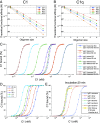
C1/C1q binding to differently sized IgG subclass oligomers. (A and B) Comparison of functional affinities of C1 (A) and C1q (B) to differently sized IgG subclass oligomers determined from kinetic simulations (until equilibrium is reached, dots) and from the thermodynamic approach (dashed lines), using the parameters from Table 2, respectively. (C) Concentration dependency of C1 binding to IgG1 monomers to hexamers at equilibrium (circles) and after an incubation time of 60 min (crosses). (D) Influence of incubation time on C1 binding to IgG1 hexamers. (E) Concentration dependency of C1 binding to IgG1 monomers to hexamers after an incubation time of 20 min. Comparison to CDC data of anti-CD20 antibody IgG1-7D8-E430G bound to Z138 cells taken from (28).
Fig. 8.
Fig. 8.
Complement-mediated lysis of DNP-coated liposomes induced by anti-DNP IgG variants. (AD) The ability of graded amounts of IgG1-4 variants to induce activation of complement in human serum is assessed by the magnitude of complement-mediated liposome lysis expressed as the change in absorbance at 340 nm (Abs) per second (dAbs/dt). Each data point represents means ± SD from n = 3 independent experiments. Lines are fits of dose–response curves to the data; EC50 values are displayed in Table 3.

References

    1. Diebolder C. A., et al. , Complement is activated by IgG Hexamers assembled at the cell surface. Science 343, 1260–1263 (2014). - PMC - PubMed
    1. Ugurlar D., et al. , Structures of C1-IgG1 provide insights into how danger pattern recognition activates complement. Science 359, 794–797 (2018). - PubMed
    1. Sharp T. H., et al. , Insights into IgM-mediated complement activation based on in situ structures of IgM-C1-C4b. Proc. Natl. Acad. Sci. U.S.A. 116, 11900–11905 (2019). - PMC - PubMed
    1. Vidarsson G., Dekkers G., Rispens T., IgG subclasses and allotypes: From structure to effector functions. Front. Immunol. 5 (2014). - PMC - PubMed
    1. Schumaker V. N., Calcott M. A., Spiegelberg H. L., Mueller-Eberhard H. J., Ultracentrifuge studies of the binding of IgG of different subclasses to the Clq subunit of the first component of complement. Biochemistry 15, 5175–5181 (1976). - PubMed

LinkOut - more resources