. 2024 Oct 22;134(24):e180282.
doi: 10.1172/JCI180282.

Sialylated glycoproteins suppress immune cell killing by binding to Siglec-7 and Siglec-9 in prostate cancer

Affiliations

Sialylated glycoproteins suppress immune cell killing by binding to Siglec-7 and Siglec-9 in prostate cancer

Ru M Wen et al. J Clin Invest. .

Abstract

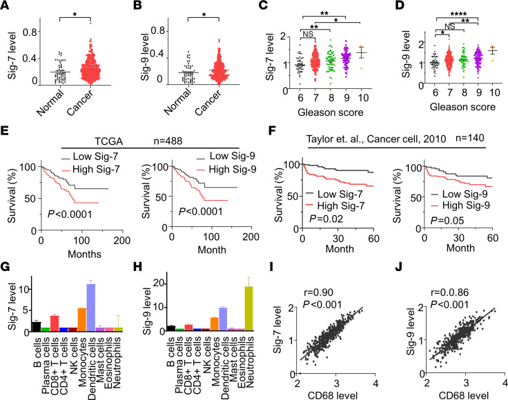
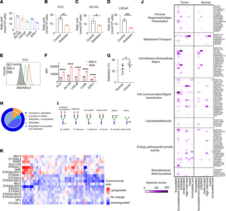
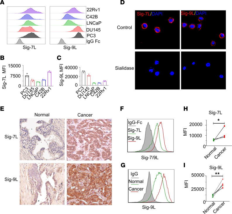
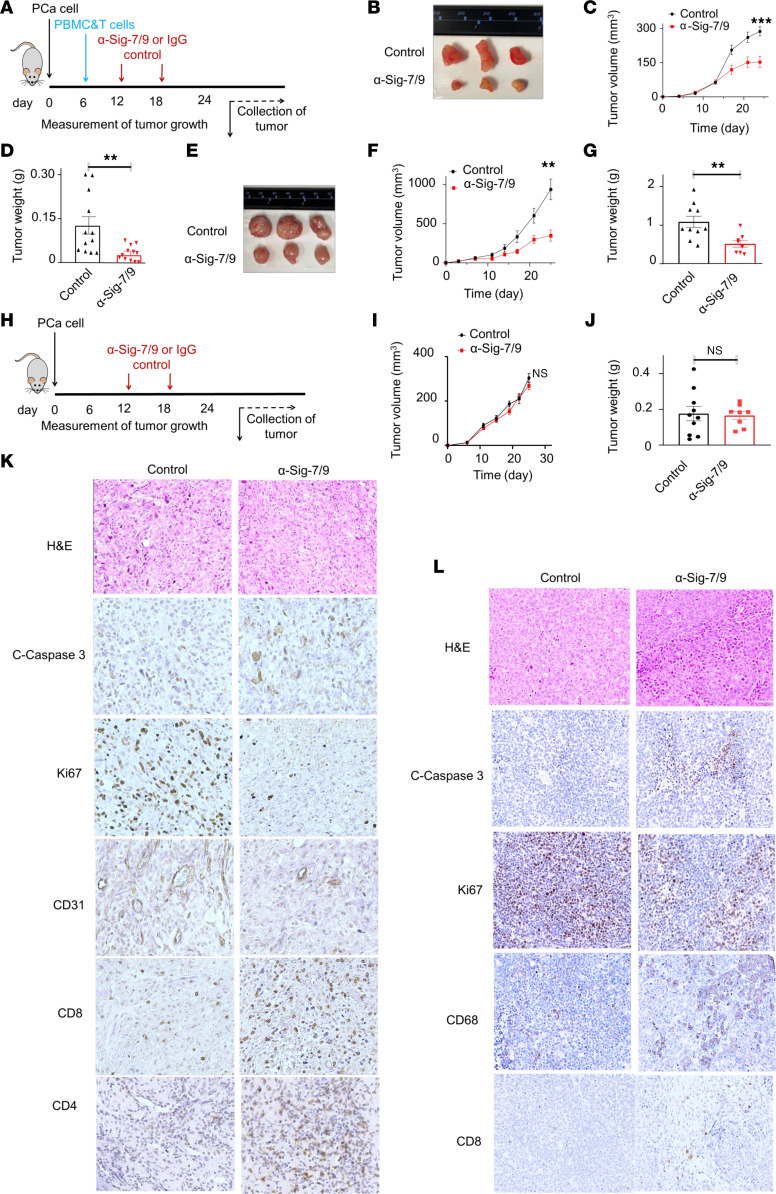
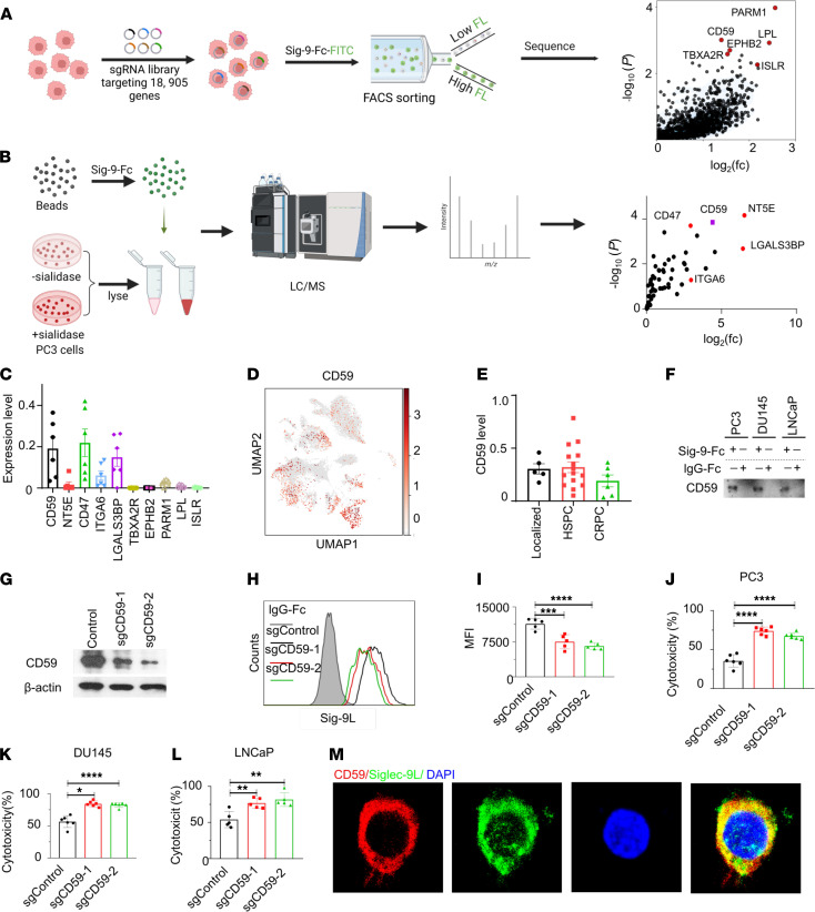
Prostate cancer is the second leading cause of male cancer death in the U.S. Current immune checkpoint inhibitor-based immunotherapies have improved survival for many malignancies; however, they have failed to prolong survival for prostate cancer. Siglecs (sialic acid-binding immunoglobulin-like lectins) are expressed on immune cells and regulate their function. Siglec-7 and Siglec-9 contribute to immune evasion in cancer by interacting with sialic acid-containing glycoprotein ligands on cancer cells. However, the role of Siglec-7/9 receptors and their ligands in prostate cancer remains poorly understood. Here, we find that Siglec-7 and Siglec-9 are associated with poor prognosis in patients with prostate cancer and are highly expressed in myeloid cells, including macrophages, in prostate tumor tissues. Siglec-7 and -9 ligands were expressed in prostate cancer cells and human prostate tumor tissues. Blocking the interactions between Siglec-7/9 and sialic acids inhibited prostate cancer xenograft growth and increased immune cell infiltration in humanized mice in vivo. Using a CRISPRi screen and mass spectrometry, we identified CD59 as a candidate Siglec-9 ligand in prostate cancer. The identification of Siglec-7 and -9 as potential therapeutic targets, including the CD59/Siglec-9 axis, opens up opportunities for immune-based interventions in prostate cancer.

Keywords: Cancer immunotherapy; Immunotherapy; Oncology; Prostate cancer.


Conflict of interest statement

Conflict of interest: RMW received a travel award from Bon Opus Biosciences. JCS has received compensation for consulting and Scientific Advisory Board membership from Rondo Therapeutics, AbTherx, and Curie Bio, as well as speaker’s honoraria from Cullinan Therapeutics, and Merck & Co. CRB is a cofounder and Scientific Advisory Board member of Lycia Therapeutics, Palleon Pharmaceuticals, Enable Bioscience, OliLux Bio, InterVenn Bio, Grace Science LLC, Firefly Bio, Redwood Bioscience (a subsidiary of Catalent), Neuravid Therapeutics, and ReNAgade Bio. JCS and CRB are cofounders of Valora Therapeutics. CRB is a member of the Board of Directors of Alnylam and OmniAb.

Figures

Figure 1
Figure 1. High Siglec-7/9 expression is correlated with worse clinical outcome in patients with PCa.
Violin plots showing that (A) Siglec-7 and (B) Siglec-9 mRNA expression are significantly higher in tumor tissues (n = 497) than normal tissues (n = 53). Data were analyzed by unpaired student’s t test. (C) Siglec-7, and (D) Siglec-9 are correlated with Gleason Score. Data were analyzed by 1-way ANOVA with post hoc Tukey’s test and presented as mean ± SEM. (E) High Siglec-7 and Siglec-9 expression is correlated with worse survival in patients with PCa in the TCGA-PRAD database (n = 488). (F) High Siglec-7 and Siglec-9 expression are correlated with worse survival in patients with PCa in MSKCC database (n = 140). Survival analysis was conducted using the Kaplan–Meier (log-rank test). Median expression was used as cutoff between high and low Siglec-7 and -9 expressing groups. (G) Siglec-7, and (H) Siglec-9 are highly expressed in monocyte, dendritic cells, and CD8+ T cells identified by CIBERSORTx from the TCGA-PRAD database. (I and J) Scatter plot of RNA expression of (I) Siglec-7 and (J) Siglec-9 compared with CD68 in prostate tumor tissues (n = 494), according to TCGA data. Siglec-7, and Siglec-9 are correlated with macrophage marker CD68. Data were analyzed by Pearson’s correlation. *P ≤ 0.05; **P ≤ 0.01; ***P ≤ 0.0001.
Figure 2
Figure 2. Siglec-7 and Siglec-9 are coexpressed on myeloid cells in human prostate tumors by single-cell RNA-seq.
(A) UMAP plot showing the distribution of cell types. (B) UMAP profiles highlighting Siglec-7 expression, and (C) Siglec-9 expression in immune cells of CRPC tumor tissues (n = 6). (D) Siglec-7 is predominantly expressed in dendritic cells (DCs), macrophages, myeloid-derived suppressor cells (MDSCs), and natural killer (NK) cells in CRPC. (E) Siglec-9 is primarily expressed in macrophages and MDSCs in CRPC specimens. (F) Siglec-7 and (G) Siglec-9 are expressed in macrophages in human tumor tissues from patients with localized PCa (n = 5), metastatic HSPC (n = 14), and metastatic CRPC (n = 6). (H) Dot plot illustrates fractional profiles of Siglec expression in immune cells across localized PCa (n = 5), metastatic HSPC (n = 14), and metastatic CRPC tumor tissues (n = 6). (I) Confocal microscopy images of a PCa bone metastasis showing the coexpression of Siglec-7 and Siglec-9 on macrophages, observed at 40 × magnification. (J) Colocalization coefficient of Siglec-7 or Siglec-9 with the macrophage marker CD68 (n = 7).
Figure 3
Figure 3. Sialic acid is expressed on the surface of PCa cells.
(A) surface sialic acid is detected in tested PCa cell lines. Sialidase treatment reduces the surface sialic acid levels in (B) PC3, (C) DU145, (D) LNCaP. (E) The expression levels of α2,6-linked and α2,3-linked sialic acids in PC3 cells were analyzed by flow cytometry using Sambucus nigra agglutinin (SNA) and Maackia amurensis agglutinin II (MALII) lectins, respectively. (F) Quantification was assessed using MFI for SNA and MALII staining on PCa cells. (G) Sialylated spectral counts versus total spectral counts of PCa tumor tissues and adjacent normal tissues by mass spectrometry. (H) Summary of glycosylation changes by glycosite in PCa compared with normal prostate samples. (I) Schematics of glycan structures. The schematic diagram was created with Biorender.com. (J) GO term pathway analysis of sialylated proteins in PCa tumor tissues and adjacent normal tissues by mass spectrometry show enrichment in immune response/antigen presentation pathways. Protein names displayed on the right side with the glycosites in parentheses. (K) Analysis of transcript data from TCGA-PRAD database of sialyglycan gene expression demonstrates 7 significantly upregulated genes and 12 downregulated genes in cancer samples compared with noncancerous prostate tissues. Data were analyzed by unpaired Student’s t test and presented as mean ± SEM. *P ≤ 0.05; **P ≤ 0.01; ***P ≤ 0.001; ***P ≤ 0.0001.
Figure 4
Figure 4. Siglec-7 ligand and Siglec-9 ligand are expressed in PCa cells.
(A) The expression levels of Siglec-7 ligand (Siglec-7L) and Siglec-9L, and (B and C) their corresponding quantification by MFI by flow cytometry. (D) Confocal microscopy analysis of PC3 PCa cells showing cell surface–associated expression patterns of Siglec-7 and Siglec-9. Treatment with sialidase abolishes the binding of Siglec-7-Fc and Siglec-9-Fc proteins visualized by confocal microscopy, indicating dependence on sialic acid. (E) Representative IHC analysis of Siglec-7 and Siglec-9 ligand expression in PCa tumor tissues. High expression levels of Siglec-7 and Siglec-9 ligands are observed in tumor tissues from patients with PCa, while normal tissue samples show either absence or low expression levels.Magnification, × 40. (F) Representative Siglec-7 and Siglec-9L expression profile on tumor cells derived from patient tumor tissues by flow cytometry, (G) Expression of Siglec-9L expression profile in all cells from tumor tissue, and adjacent normal tissues (n = 6). Quantification of (H) Siglec-7L and (I) Siglec-9L in samples from patients with PCa, suggesting significant higher expression levels of Siglec-7L and Siglec-9L in tumor tissues compared with adjacent normal tissues. Data were analyzed paired Student’s t test and presented as mean ± SEM.; *P ≤ 0.05; **P ≤ 0.01.
Figure 5
Figure 5. Siglec-7/9 receptor blockade restricts PCa tumor growth in humanized mouse models.
(A) Schematic diagram depicting the implantation of PCa cells into NSG mice, followed by injection of PBMC and CD8+ T cell mixture and subsequent administration of anti-Siglec-7 and anti-Siglec-9 mAbs or IgG antibodies on specific days. (B) Representative images of PC3 tumors upon treatment with anti-Siglec-7/9 antibodies (n = 12) compared with IgG control (n = 12). (C) PC3 tumor growth curve and corresponding (D) tumor weight. (E) Representative images of 22Rv1 tumors upon treatment with anti-Siglec-7/9 antibodies (n = 8) compared with IgG control (n = 10). (F) 22Rv1 tumor growth curve, and corresponding (G) tumor weight. (H) Schematic diagram depicting control experiment without PBMC and CD8+ T cell injection. Control experiment no difference in (I) PC3 tumor growth and (J) tumor weight between anti-Siglec-7/9 antibody (n = 8) and IgG1 isotype control treatment (n = 10). (K) IHC staining of PC3 tumors treated with anti-Siglec-7/9 antibodies showing increased apoptosis (cleaved caspase 3), decreased proliferation (Ki67), decreased vascularization (CD31), and increased immune cell infiltration, including CD4+ and CD8+ T cells, compared with IgG1 control. Magnification, × 40. (L) IHC staining of 22Rv1 tumors treated with anti-Siglec-7/9 antibodies showing increased apoptosis (cleaved caspase 3), decreased proliferation (Ki67), decreased vascularization (CD31), increased macrophage (CD68) infiltration, and enhanced CD8+ T cell numbers compared with IgG1 control. Data were analyzed by unpaired Student’s t test and presented as mean ± SEM. **P ≤ 0.01; ***P ≤ 0.001.
Figure 6
Figure 6. CD59 is a Siglec-9 ligand in PCa.
(A) Experimental setup and CRISPRi screen results for identifying Siglec-9 ligands using CRISPR screen. PC3 cells expressing dCas9 were infected with a genome-wide CRISPRi-v2 library, and cells with reduced binding to recombinant Siglec-9 Fc proteins were sorted for deep sequencing (n = 3). The schematic diagram was created with Biorender.com. (B) Identification of sialic acid–dependent Siglec-9 ligands through LC-MS/MS pull down analysis by Siglec-9 Fc chimera protein (n = 3). The schematic diagram was created with Biorender.com. (C) Analysis of transcript levels of Siglec-9 ligands by sc-RNA sequencing. CD59 and candidate ligands identified by CRISPRi and MS expressed in metastatic epithelial cells from patients with CRPC. (D) UMAP profile shows the distribution of CD59 in different cell types in human metastatic CRPC tumor tissues, refer to Figure 2A for cell type distribution. (E) CD59 is expressed in cancer epithelial cells from patients with localized, HSPC, and CRPC. (F) Western blotting validation of CD59 as a Siglec-9 ligand using recombinant Siglec-9 Fc protein blotting pull-down blotting. (G) Western blot analysis of CD59-KO PC3 cells using the CRISPR/Cas9 system. (H) Flow cytometry analysis and (I) corresponding quantification demonstrating reduced binding capacity of Siglec-9 Fc in CD59 knockout PC3 cells compared with control cells. Gray, IgG Fc; black, control cells; red, sgCD59-T1; green, sgCD59-T2. (J) Enhanced cytotoxicity of CD8+ T cells were observed in CD59-KO PC3 cells, (K) DU145 cells, and (L) LNCaP cells. (M) Confocal microscopy showing substantial colocalization between Siglec-9 ligands and CD59 on PC3 cells. Data were analyzed by 1-way ANOVA with post hoc Tukey’s test and presented as mean ± SEM.*P ≤ 0.05; **P ≤ 0.01; ***P ≤ 0.001; ***P ≤ 0.0001.

References

    1. Siegel RL, et al. Cancer statistics 2023. CA Cancer J Clin. 2023;73(1):17–48. doi: 10.3322/caac.21763. - DOI - PubMed
    1. Lowrance WT, et al. Castration-resistant prostate cancer: AUA Guideline Amendment 2015. J Urol. 2016;195(5):1444–1452. doi: 10.1016/j.juro.2015.10.086. - DOI - PubMed
    1. Shou J, et al. The prognosis of different distant metastases pattern in prostate cancer: A population based retrospective study. Prostate. 2018;78(7):491–497. doi: 10.1002/pros.23492. - DOI - PubMed
    1. Naimi A, et al. Tumor immunotherapies by immune checkpoint inhibitors (ICIs); the pros and cons. Cell Commun Signal. 2022;20(1):44. doi: 10.1186/s12964-022-00854-y. - DOI - PMC - PubMed
    1. Robert C. A decade of immune-checkpoint inhibitors in cancer therapy. Nat Commun. 2020;11(1):3801. doi: 10.1038/s41467-020-17670-y. - DOI - PMC - PubMed

MeSH terms