. 2024 Dec;11(46):e2405823.
doi: 10.1002/advs.202405823. Epub 2024 Oct 22.

A Programmable Handheld Extrusion-Based Bioprinting Platform for In Situ Skin Wounds Dressing: Balance Mobility and Customizability

Affiliations

A Programmable Handheld Extrusion-Based Bioprinting Platform for In Situ Skin Wounds Dressing: Balance Mobility and Customizability

Chenmin Wang et al. Adv Sci (Weinh). 2024 Dec.

Abstract

Bioprinting technology plays a crucial role for constructing tissue substitutes. However, the mismatched scaffold shapes and the poor treatment timeliness limit its clinical translational application. In situ printing technology that prints bioregenerants directly inside patient's body can meet the needs of specific tissue repair. This study develops a smartphone controlled handheld bioprinter for in situ skin wounds dressing. The mini bioprinter can be handheld and placed on any printing surface to create strips, complex patterns, and 3D structures, and can be equipped with microchannel needles to expand functionality. The size of the strips as well as the printing path can be programmed and controlled by the smartphone to ensure the precision of the printed product quality. Furthermore, the device not only allows for smooth switching between different bioinks for printing heterogeneous structure, but also allows for fast and uniform coverage of large wound surfaces. When dealing with complex wounds in vitro & vivo, the printer can effectively fill and precisely close wounds, promoting wound healing. The programmable handheld bioprinter can balance mobility and customizability in the management of skin wounds and is expected to realize its potential for emergency medical treatment in condition-constrained scenarios, such as battlefields or disaster areas.

Keywords: handheld printers; in situ bioprinting; programmable; skin regeneration.


Conflict of interest statement

The authors declare no conflict of interest.

Figures

Figure 1
Figure 1
Schematic representation of the concept and structure of in situ bioprinting equipment. A) a robotic arm system under digital program guidance and B) a handheld printing device manually deposit bioinks onto tissue defects to perform in situ bioprinting. C) Illustration of the programmable handheld bioprinter dispensing bioinks in a controlled, uniform manner as directed by programmed instructions while being operated manually to fill tissue defects as needed. D) Photographs of the programmable handheld bioprinter from different orientations (The square on the table mat has a side length of 5 cm).
Figure 2
Figure 2
Illustration of the operational workflow of the programmable handheld printer. From left to right, the process initiates with a smartphone connecting to and controlling the printer via Wi‐Fi. This is followed by the selection of either a pneumatic or mechanical extrusion method; setting the corresponding printing parameters; and loading the bioink. Subsequently, the user selects the desired mode in the Web App's printing path planning interface, which generates the program instructions for printing. Finally, the printer is manually operated to execute the printing task.
Figure 3
Figure 3
Rheological evaluation of two bioink systems. A–C) depict the shear‐thinning behavior of the laponite XLG‐based bioink system under shear rates ranging from 0.1 to 100 s−1, the oscillatory shear test across shear strains from 1% to 100%, and the temperature sensitivity evaluation within the range of 4 to 40 °C, respectively. D–F) correspond to the granular hydrogel bioink system, presenting the same tests for shear‐thinning behavior, oscillatory shear tests, and temperature sensitivity evaluation under identical testing conditions.
Figure 4
Figure 4
The performance of the strips printing by the programmable handheld bioprinter. A–D) Strip width variations with Laponite XLG‐based bioink compositions, nozzle sizes, extrusion rates and printing speed index under mechanical extrusion mode; scale bar = 500 µm. E) Mass of Laponite XLG‐based bioinks extruded per 60 s at various extrusion rates under mechanical extrusion mode, and its linear fitting was performed. F–I) Strip width variations with granular hydrogel bioink compositions, nozzle sizes, extrusion pressure index, and printing speed index under pneumatic extrusion mode; scale bar = 5 mm. J) Mass of granular hydrogel bioinks extruded per 60 s at various extrusion pressures index under pneumatic mode, and its linear fitting was performed. (Notes: The shared control group is highlighted in red. !: Discontinuous strips were excluded from the statistical analysis. p < 0.05 (*), p < 0.01 (**), and p < 0.001 (***)).
Figure 5
Figure 5
Demonstration of programmable handheld printing for mono/multi‐bioink distribution of voxelization. A) Pixelated “S”, “I”, “A”, “T” characters were input into the Web App's Self‐Designed path planning interface to generate corresponding program instructions, directing the programmable handheld printer to print granular hydrogel bioinks and form a monochrome pattern. B–E) Building upon the mono‐bioink pattern, color images were broken down into multiple printing channels by color and programmed individually. Sequential programmable handheld printing of each channel's granular hydrogel bioink formed complex smiley face and fruit patterns composed of multiple bioinks. F) Using Laponite XLG‐based bioink and manual adjustment of the programmable handheld printer's position facilitated the printing of mono‐bioink cubes (I) and pyramids (II), as well as bi‐bioink cubes (III), and pyramids (IV).
Figure 6
Figure 6
Printing with microchannel nozzles on the programmable handheld printer. A) Displayed from top to bottom the 3D rendering, actual image, and the nozzle attached to the printer for the “all‐in‐one” microchannel nozzle. B,C) Illustrated the bioinks distribution within the “all‐in‐one” microchannel nozzle during the printing of mono and multi‐bioink, with corresponding photographs of the resulting strips. The transparent color represented granular hydrogel bioinks extruded pneumatically, while other colors indicated laponite XLG‐based bioinks extruded mechanically. The dot plots in (C) indicated the extrusion rates of different colored bioinks during printing of various rows of patterns. Scale bar = 1.00 cm. D) Displayed from top to bottom the 3D rendering, actual image, and the nozzle attached to the printer for the “thin‐layer” microchannel nozzle. E,F) Showed the bioinks distribution diagrams within the “thin‐layer” microchannel nozzle during mechanical extrusion of single and double layers of laponite XLG‐based bioinks, along with photographs of the corresponding thin layers produced. Scale bar = 1.00 cm.
Figure 7
Figure 7
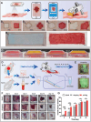
Programmable handheld in situ bioprinting for skin wound treatment. A) Schematic of the process for programmable handheld in situ bioprinting on in vitro porcine skin, including steps for wound image capture, gridding of the image, generation of printing program instructions, and bioprinting the granular hydrogel bioink to cover the wound B) Before and after images of programmable handheld in situ bioprinting on in vitro porcine skin wounds in various shapes: square (B1), cross (B2), hexagon (B3), random (B4), and a large‐area rectangle (B5). Scale bar = 1.00 cm. C). The programmable handheld bioprinter with a “thin‐layer” microchannel nozzle was used to print four layers of 3@10 bioink onto the simulated human full‐thickness skin wound model, effectively closing the “wound”. Different colored pigments were added between adjacent layers of bioink to distinguish each layer visually. D) Illustration of the in situ bioprinting process for a rat full‐thickness skin wound model using the “thin‐layer” microchannel nozzle, including preparing bioinks (PRP mixed with microskin grafts), in situ bioprinting of the bioinks, and post‐surgical care and observation of the rats. E) Comparison of bioinks distribution on rat skin wounds using droplet method versus programmable handheld printing, with green fluorescent dye added to the bioinks for visibility. F) Photographs depicting the healing status of rat skin wounds treated with different methods; scale bar = 1.00 cm, with wound closure rates quantified in G). (p < 0.05 (*), p < 0.01 (**), and p < 0.001 (***)).

References

    1. a) Fonseca A. C., Melchels F. P. W., Ferreira M. J. S., Moxon S. R., Potjewyd G., Dargaville T. R., Kimber S. J., Domingos M., Chem. Rev. 2020, 120, 11093; - PMC - PubMed
    2. b) Wang X., Luo Y., Ma Y., Wang P., Yao R., Trends Biotechnol. 2023, 42, 648; - PubMed
    3. c) Daly A. C., Prendergast M. E., Hughes A. J., Burdick J. A., Cell 2021, 184, 18. - PMC - PubMed
    1. You S. T., Xiang Y., Hwang H. H., Berry D. B., Kiratitanaporn W., Guan J. A., Yao E., Tang M., Zhong Z., Ma X. Y., Wangpraseurt D., Sun Y. Z., Lu T. Y., Chen S. C., Sci. Adv. 2023, 9, ade7923. - PMC - PubMed
    1. a) O'Connor C., Brady E., Zheng Y., Moore E., Stevens K. R., Nat. Rev. Mater. 2022, 7, 702; - PMC - PubMed
    2. b) Wang Z. J., Liang X., Wang G. Y., Wang X. H., Chen Y., Adv. Mater. 2023, 2304738;
    3. c) Kang H. W., Lee S. J., Ko I. K., Kengla C., Yoo J. J., Atala A., Nat. Biotechnol. 2016, 34, 312; - PubMed
    4. d) de Melo B. A. G., Jodat Y. A., Mehrotra S., Calabrese M. A., Kamperman T., Mandal B. B., Santana M. H. A., Alsberg E., Leijten J., Shin S. R., Adv. Funct. Mater. 2019, 29, 1906330; - PMC - PubMed
    5. e) Wang C. M., Honiball J. R., Lin J. Y., Xia X. Y., Lau D. S. A., Chen B., Deng L. F., Lu W. W., ACS Appl. Mater. Interfaces 2022, 14, 27575. - PubMed
    1. a) Vijayavenkataraman S., Yan W. C., Lu W. F., Wang C. H., Fuh J. Y. H., Adv. Drug Delivery Rev. 2018, 132, 296; - PubMed
    2. b) Samandari M., Mostafavi A., Quint J., Memić A., Tamayol A., Trends Biotechnol. 2022, 40, 1229. - PMC - PubMed
    1. Nuutila K., Samandari M., Endo Y., Zhang Y. T., Quint J., Schmidt T. A., Tamayol A., Sinha I., Bioact. Mater. 2022, 8, 296. - PMC - PubMed

LinkOut - more resources