. 2024 Nov:453:109134.
doi: 10.1016/j.heares.2024.109134. Epub 2024 Oct 18.

Critical role of hepsin/TMPRSS1 in hearing and tectorial membrane morphogenesis: Insights from transgenic mouse models

Affiliations

Critical role of hepsin/TMPRSS1 in hearing and tectorial membrane morphogenesis: Insights from transgenic mouse models

Ting-Hua Yang et al. Hear Res. 2024 Nov.

Abstract

Mutations in various type II transmembrane serine protease (TMPRSS) family members are associated with non-syndromic hearing loss, with some mechanisms still unclear. For instance, the mechanism underlying profound hearing loss and tectorial membrane (TM) malformations in hepsin/TMPRSS1 knockout (KO) mice remains elusive. In this study, we confirmed significantly elevated hearing thresholds and abnormal TM morphology in hepsin KO mice, characterized by enlarged TM with gaps and detachment from the spiral limbus. Transgenic mouse lines were created to express either wild-type or a serine protease-dead mutant of human hepsin in the KO background. The Tg68;KO line, expressing moderate levels of wild-type human hepsin in the cochlea, showed partial restoration of hearing function. Conversely, the Tg5;KO or TgRS;KO lines, with undetectable hepsin or protease-dead hepsin, did not show such improvement. Histological analyses revealed that Tg68;KO mice, but not Tg5;KO or TgRS;KO mice, had a more compact TM structure, partially attached to the spiral limbus. These results indicate that hepsin expression levels correlate with improvements in hearing and TM morphology, and its protease activity is critical for these effects. Hepsin's role was further examined by studying its relationship with α-tectorin (TECTA) and β-tectorin (TECTB), non-collagenous proteins crucial for TM formation. Hepsin was co-expressed with TECTA and TECTB in the developing cochlear epithelium. Immunostaining showed decreased levels of TECTA and TECTB in hepsin KO TM, partially restored in Tg68;KO mice. These findings suggest that hepsin is essential for proper TM morphogenesis and auditory function, potentially by proteolytic processing/maturation of TECTA and TECTB and their incorporation into the TM.

Keywords: Hearing function; Hepsin; Tectorial membrane; Tectorin; Transgenic mice; Type II transmembrane serine protease.


Conflict of interest statement

Declaration of competing interest None.

Figures
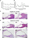
Fig. 1.
Fig. 1.. Profound hearing loss and TM malformation in hepsin KO mice.
(A) ABR tests with click stimuli and pure tone bursts at 4 kHz, 8 kHz, 16 kHz, and 32 kHz revealed that 6–8 week-old hepsin knockout (KO) mice had significantly higher ABR thresholds compared to their wild type (WT) littermate controls, indicating poorer hearing ability in the KO mice (two-way ANOVA, n = 20 ears from 10 mice per group, p<0.0001). ABR thresholds were defined as the lowest sound pressure level at which reproducible waveforms were detectable. (B) DPOAE amplitudes were plotted after subtracting the background noise from the measured values. While DPOAEs were detectable in both WT and hepsin KO mice, the amplitudes above 12 kHz were reduced in KO mice compared to WT controls [two-way ANOVA, WT vs. KO, n = 20 ears from 10 mice per group, F(1,38) = 23.50, p < 0.0001]. (C and D) H&E staining of cochlear sections from adult WT and hepsin KO mice revealed structural abnormalities in the TM of KO mice. Specifically, sections from the middle turn (16 kHz region) and apical turn (8 kHz region) of KO mice displayed pronounced enlargement and the presence of abnormal spaces within the TM compared to WT controls. Additionally, the TM was detached from the spiral limbus in KO mice. (E) H&E staining of cochlear sections from P1 WT and hepsin KO mice revealed TM malformations in the KO mice at this early stage. However, compared to adult KO mice, the TM enlargement and detachment were less pronounced at P1. Scale bar: 50 μm (C and D), 100 μm (E).
Fig. 2.
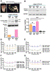
Fig. 2.. Improvement of hearing in hepsin KO mice by expression of wild type human hepsin transgene in the organ of Corti.
(A) Gross image of an isolated murine inner ear from an adult mouse (left). The organ of Corti can be separated from the stria vascularis in the dissected inner ear (right). (B) RT-PCR analysis of human hepsin expression in the organ of Corti from 6- to 8-week-old mice. Mouse groups include WT, hepsin KO, and three transgenic lines expressing human hepsin in the hepsin KO background (Tg68;KO, Tg5;KO, TgRS;KO). The PCR-amplified hepsin DNA fragment is 300 bp. The bottom plot shows the expression ratio of human hepsin, normalized to endogenous GAPDH as an internal control, based on RT-PCR assays performed in triplicate. TgRS;KO mice expressed the highest level of human hepsin, while Tg5;KO mice expressed the lowest. (C) Western blot analysis of protein extracts from the organ of Corti collected from 12 adult mice. The bottom plot shows the expression ratio of human hepsin protein, normalized to endogenous β-actin levels as an internal control. TgRS;KO mice showed high expression levels of human hepsin, while Tg68;KO mice exhibited low expression. Human hepsin protein was absent in the organ of Corti from KO mice. (D-F) Auditory thresholds of various 6–8 week-old transgenic mouse lines and their littermate controls were measured by ABR tests. Breeding pairs were set up as Tg+;HPN+/− crossed with Tg-;HPN+/− for all three transgenic lines to generate different combinations of littermate controls for Tg+;KO mice. Auditory thresholds were normal in WT and hepsin heterozygous (Het) littermate controls from the Tg-hHPN breeding (including all Tg;WT and Tg;Het mice). Hepsin KO littermates had significantly higher auditory thresholds at all stimulus frequencies. However, among the three transgenic lines (Tg68, Tg5, and TgRS), the auditory thresholds of Tg68;KO mice were significantly improved upon expressing wild type human hepsin in the organ of Corti (two-way ANOVA, Tg68-;KO vs. Tg68+;KO, p<0.0001). In contrast, the absence of detectable human hepsin protein expression in Tg5;KO cochlea or expression of the protease-dead mutant of human hepsin in TgRS;KO mice did not improve the hearing threshold in hepsin KO mice (two-way ANOVA, Tg5-;KO vs. Tg5+;KO, p=0.4694; TgRS-;KO vs. TgRS+;KO, p=0.3178). (G) Auditory thresholds of the three Tg-hHPN mice in the hepsin KO background. Compared to Tg5;KO or TgRS;KO mice, Tg68;KO mice showed significant improvement in hearing thresholds at all stimulus frequencies (two-way ANOVA, Tg5;KO vs. Tg68;KO, p<0.0001; TgRS;KO vs. Tg68;KO, p<0.0001).
Fig. 3.
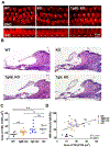
Fig. 3.. Morphological analysis of auditory hair cells and the TM.
(A) Confocal microscope images of inner and outer hair cells. Whole-mount organ of Corti was stained with Phalloidin and imaged using a confocal microscope. The morphology of inner and outer hair cells appeared normal among adult WT (Tg68-;HPN+/+), KO (Tg68-;HPN−/−), and Tg68;KO (Tg68+;HPN−/−) littermate mice. Inner hair cells (IHC) and outer hair cells (OHC) are indicated, with white arrows pointing to the hair bundles. Scale bar, 10 μm. (B) H&E staining of cochlear sections from 6- to 8-week-old WT, KO, Tg68;KO and Tg5;KO mice. The TM in Tg5;KO mice was enlarged and detached from the spiral limbus, similar to the TM in hepsin KO mice. In contrast, the TM in Tg68;KO mice appeared thinner, more compact, less detached from the spiral limbus, and more closely resembled that of WT mice. (C) Quantification of TM sectional area using Axovision software. The average TM sectional areas (μm2) were 1556.03 ± 459.50 (n=8 ears) for WT, 2175.39 ± 567.75 (n=13 ears) for Tg68;KO, 3092.55 ± 1284.62 (n=11 ears) for Tg5;KO, and 3227.94 ± 910.61 (n=11 ears) for KO mice. Statistical analysis was performed using one-way ANOVA followed by post hoc Tukey’s test: *p<0.05, **p<0.01, ns: not significant. Specific comparisons yielded the following p-values: WT vs. Tg68;KO, p = 0.4084; WT vs. Tg5;KO, p = 0.003; WT vs. KO, p = 0.0011; Tg68;KO vs. Tg5;KO, p = 0.0682; Tg68;KO vs. KO, p = 0.0282; and Tg5;KO vs. KO, p = 0.9836. (D) Linear regression analysis of the TM sectional area and ABR thresholds for click responses from WT, Tg68;KO, Tg5;KO, and KO mice. ABR thresholds were determined as the lowest sound pressure level at which reproducible waveforms were detectable. The analysis yielded an R2 value of 0.1751, with a significance of p=0.0052.
Fig. 4.
Fig. 4.. Ultrastructural analysis of the TM by transmission electron microscopy.
Electron micrographs of the TM ultrastructure from the mid-modiolar (16-kHz) region of 6- to 8-week-old WT, KO, Tg68;KO, Tg5;KO, and TgRS;KO cochleae. The mouse TM is composed of collagens, which constitute approximately 50% of total TM proteins and are radially oriented as collagen-fibril bundles. In WT TM, the collagen bundles are dense, continuous, and uniformly spaced in parallel. The arrangement of collagen fibrils in Tg68;KO TM closely resembles that of WT TM. In contrast, the collagen bundles in the TMs of KO, Tg5;KO, and TgRS;KO mice appear discontinuous, fragmented, and loosely organized. Magnification at 51200X. Scale bar: 500 nm. The area of the TM occupied by collagen fibers was calculated using ImageJ, divided by the total area of the micrograph, and presented as percentages (n = 2 to 3 fields at 51,200× per ear, with 2 ears per group). One-way ANOVA, *p<0.05, **p<0.01. Scale bar: 500 nm.
Fig. 5.
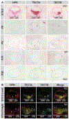
Fig. 5.. Hepsin is co-expressed with TECTA and TECTB in the GER and LER of the developing cochlear duct.
(A) The expression patterns of hepsin (HPN), α-tectorin (TECTA), and β-tectorin (TECTB) in mouse cochleae were detected by RNAScope in situ hybridization using a horseradish peroxidase (HRP)-based chromogen (red) at E15.5, P0, P5, P10, and P20. Cochleae were outlined with dashed lines based on DAPI counterstaining. Hepsin expression is evident in both the GER and LER at E15.5 but becomes downregulated at early postnatal stages. TECTA exhibits high expression levels in the GER and LER in embryonic and early postnatal stages (as indicated by arrows), progressively downregulating and becoming undetectable after P10. TECTB shows expression in two distinct zones within both the GER and LER from the embryonic stage up to P10 (as indicated by arrows), gradually decreasing and becoming undetectable after P15. The sensory epithelium, marked by asterisks, is shown at high magnification in the inset. (B) The expression patterns of HPN, TECTA, and TECTB in an E15.5 mouse cochlear duct were simultaneously detected by RNAScope multiplex in situ hybridization using HRP-based TSA Vivid fluorophores [HPN: TSA Vivid 570 (red), TECTA: TSA Vivid 650 (pseudo-colored yellow), and TECTB: TSA Vivid 520 (green)]. The lower panels are high-magnification images corresponding to those in the upper panels. Hepsin expression was found to co-localize with both TECTA and TECTB expression within both the GER and LER (as indicated by arrows). Scale bar: 200μm (E15.5), 400μm (P0-P20 in A), 80μm (insets), 74μm (upper panels in B), 33μm (lower panels in B).
Fig. 6.
Fig. 6.. Analysis of collagenous and non-collagenous protein components in the TM using immunofluorescence staining.
(A) Fixed frozen sections from the mid-modiolar (16-kHz) region of 6–8 week-old WT, Tg68;KO, Tg5;KO, TgRS;KO, and KO cochleae were immunostained with antibodies against α-tectorin (left row, red), β-tectorin (middle row, green), and type II collagen (right row, red). Images were acquired using Zeiss confocal microscopy. In the Tg68;KO TM, α-tectorin and β-tectorin staining is increased compared to KO littermates (Tg68-;HPN−/−) and is nearly as strong as in WT littermate controls (Tg68-; HPN+/+). In KO mice, α-tectorin staining is weaker in the upper part of the TM and at the connection with the spiral limbus. Notable signals of β-tectorin expression in the KO TM are mainly observed at the tip and lower half of the TM. Immunostaining of type II collagen showed strong intensity covering almost the entire TM. Scale bar: 50 μm. (B) Quantification of immunofluorescence intensity of α-tectorin, β-tectorin, and type II collagen in the TM. The intensity in the TM was normalized by the total TM area indicated based on Hoechst counterstaining. The number of dots denotes the sample size of cochleae from each group of mice. One-way ANOVA, *p<0.05, **p<0.01.

References

    1. Brunati M, Perucca S, Han L, Cattaneo A, Consolato F, Andolfo A, Schaeffer C, Olinger E, Peng J, Santambrogio S, Perrier R, Li S, Bokhove M, Bachi A, Hummler E, Devuyst O, Wu Q, Jovine L, Rampoldi L, 2015. The serine protease hepsin mediates urinary secretion and polymerisation of Zona Pellucida domain protein uromodulin. Elife 4, e08887. 10.7554/eLife.08887. - DOI - PMC - PubMed
    1. Coutinho P, Goodyear R, Legan PK, Richardson GP, 1999. Chick alpha-tectorin: molecular cloning and expression during embryogenesis. Hear Res 130(1–2), 62–74. 10.1016/s0378-5955(98)00213-5. - DOI - PubMed
    1. Garrick D, Fiering S, Martin DI, Whitelaw E, 1998. Repeat-induced gene silencing in mammals. Nat Genet 18(1), 56–59. 10.1038/ng0198-56. - DOI - PubMed
    1. Goodyear RJ, Richardson GP, 2002. Extracellular matrices associated with the apical surfaces of sensory epithelia in the inner ear: molecular and structural diversity. J Neurobiol 53(2), 212–227. 10.1002/neu.10097. - DOI - PubMed
    1. Goodyear RJ, Richardson GP, 2018. Structure, Function, and Development of the Tectorial Membrane: An Extracellular Matrix Essential for Hearing. Curr Top Dev Biol 130, 217–244. 10.1016/bs.ctdb.2018.02.006. - DOI - PubMed

MeSH terms

LinkOut - more resources