. 2024 Dec 1;327(6):L917-L929.
doi: 10.1152/ajplung.00280.2023. Epub 2024 Oct 22.

Longitudinal microcomputed tomography detects onset and progression of pulmonary fibrosis in conditional Nedd4-2 deficient mice

Affiliations

Longitudinal microcomputed tomography detects onset and progression of pulmonary fibrosis in conditional Nedd4-2 deficient mice

Dominik H W Leitz et al. Am J Physiol Lung Cell Mol Physiol. .

Abstract

Idiopathic pulmonary fibrosis (IPF) is a fatal lung disease, which is usually diagnosed late in advanced stages. Little is known about the subclinical development of IPF. We previously generated a mouse model with conditional Nedd4-2 deficiency (Nedd4-2-/-) that develops IPF-like lung disease. The aim of this study was to characterize the onset and progression of IPF-like lung disease in conditional Nedd4-2-/- mice by longitudinal micro-computed tomography (CT). In vivo micro-CT was performed longitudinally in control and conditional Nedd4-2-/- mice at 1, 2, 3, 4, and 5 mo after doxycycline induction. Furthermore, terminal in vivo micro-CT followed by pulmonary function testing and post mortem micro-CT was performed in age-matched mice. Micro-CT images were evaluated for pulmonary fibrosis using an adapted fibrosis scoring system. Histological assessment of lung collagen content was conducted as well. Micro-CT is sensitive to detect the onset and progression of pulmonary fibrosis in vivo and to quantify distinct radiological IPF-like features along disease development in conditional Nedd4-2-/- mice. Nonspecific interstitial alterations were detected from 3 mo, whereas key features such as honeycombing-like lesions were detected from 4 mo onward. Pulmonary function correlated well with in vivo (r = -0.738) and post mortem (r = -0.633) micro-CT fibrosis scores and collagen content. Longitudinal micro-CT enables in vivo monitoring of the onset and progression and detects radiological key features of IPF-like lung disease in conditional Nedd4-2-/- mice. Our data support micro-CT as a sensitive quantitative endpoint for the preclinical evaluation of novel antifibrotic strategies.NEW & NOTEWORTHY IPF diagnosis, particularly in early stages, remains challenging. In this study, micro-CT is used in conditional Nedd4-2-/- mice to closely monitor the onset and progression of progressive pulmonary fibrosis in vivo. Together with high-resolution post mortem micro-CT, this allowed us to track how nonspecific lung lesions develop into key IPF-like features. This approach offers a noninvasive method to monitor pulmonary fibrosis, providing a quantitative endpoint for the preclinical evaluation of novel antifibrotic strategies.

Keywords: IPF; Nedd4-2; animal model; idiopathic pulmonary fibrosis; micro-CT.


Conflict of interest statement

No conflicts of interest, financial or otherwise, are declared by the authors.

Figures

None
Graphical abstract
Figure 1.
Figure 1.
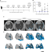
Longitudinal in vivo micro-CT detects onset and progression of pulmonary fibrosis in conditional Nedd4-2−/− mice. A: schematic illustration of the longitudinal study design with serial in vivo micro-CT 1, 2, 3, 4, and 5 mo after doxycycline induction of conditional Nedd4-2−/− mice and controls. B: summary of in vivo micro-CT fibrosis scores assessed by longitudinal micro-CT. C: representative longitudinal in vivo micro-CT image of a spontaneously breathing conditional Nedd4-2−/− mouse scanned at the indicated time points after doxycycline induction. Fibrotic areas are indicated in blue. D: 3-D reconstructions of fibrotic lesions (blue) in the lung of the same mouse as shown in C after 2, 3, 4, and 5 mo of doxycycline induction. n = 5–15 mice/group. *P < 0.05, ***P < 0.001. 3-D, three-dimensional; micro-CT, micro-computed tomography.
Figure 2.
Figure 2.
Comparison of radiological features determined by in vivo and post mortem micro-CT during the development of IPF-like lung disease in conditional Nedd4-2−/− mice. A: study design of cross-sectional micro-CT imaging during the development of IPF-like lung disease in conditional Nedd4-2−/− mice using in vivo micro-CT imaging followed by pulmonary function testing (PFT) and a post mortem micro-CT scan after 1, 2, 3, 4, and 5 mo of doxycycline induction. B and C: representative in vivo micro-CT images in spontaneously breathing conditional Nedd4-2−/− mice (B) and post mortem micro-CT (C) at indicated time points. D: Comparison of consolidations (red asterisks), honeycombing-like lesions (black arrowheads), peripheral bronchiectasis (white arrows), parenchymal lines (black arrows), and reticulations (white arrowheads) in in vivo and post mortem micro-CT 4 mo after conditional Nedd4-2 deletion. IPF, idiopathic pulmonary fibrosis; micro-CT, micro-computed tomography.
Figure 3.
Figure 3.
Development of radiological features of IPF-like lung disease in conditional Nedd4-2−/− mice. A: in vivo micro-CT fibrosis score along disease development after 1, 2, 3, 4, and 5 mo of doxycycline induction in conditional Nedd4-2−/− mice and controls. Post mortem micro-CT fibrosis score (B), consolidations (C), honeycombing-like lesions (D), fissural thickening (E), peripheral bronchiectasis (F), parenchymal lines (G), and reticulations (H) detected by high-resolution micro-CT in conditional Nedd4-2−/− mice and controls. Females are indicated as filled circles, males are indicated as open circles. n = 5–12 mice/group. FDR-adjusted P values are indicated as *P < 0.05, **P < 0.01, ***P < 0.001. IPF, idiopathic pulmonary fibrosis; micro-CT, microcomputed tomography.
Figure 4.
Figure 4.
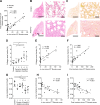
Micro-CT fibrosis score negatively correlates with impairment in pulmonary function in conditional Nedd4-2−/− mice. A: correlation between fibrosis scores determined by in vivo micro-CT and post mortem micro-CT. n = 31 mice. B: representative micrographs of H&E-stained lung sections of 4-mo-induced conditional Nedd4-2−/− and control mice. Low magnification (left, scale bar 2.5 mm). Consolidations (red asterisks), honeycombing-like lesions (black arrowheads), and peripheral bronchiectasis (white arrows). Higher magnification of cystic structural lung damages in the periphery of the lung (bottom right image, scale bar 100 µm). C: representative micrographs of histological sections stained for picrosirius red from 4-mo-induced conditional Nedd4-2−/− and control mice at low (left, scale bar 2.5 mm) and high magnifications (right, scale bar 100 µm). D: quantification of collagen in picrosirius red-stained lung sections showing the collagen area fraction in controls and conditional Nedd4-2−/− mice after the indicated periods of induction. n = 3–6 mice/group. Correlation between collagen area fraction and in vivo micro-CT fibrosis score (E) or post mortem micro-CT fibrosis score (F). n = 22 mice. G: summary of lung compliance at different time points in conditional Nedd4-2−/− mice compared with controls. n = 3–7 mice/group. Correlation between lung compliance determined by pulmonary function testing and in vivo micro-CT fibrosis score (H) (n = 35 mice) and post mortem micro-CT fibrosis score (I) (n = 37 mice) in conditional Nedd4-2−/− mice. Females are indicated as filled circles, males are indicated as open circles. *P < 0.05, **P < 0.01. Spearman rank order correlation coefficient (r), and P values are provided. H&E, hematoxylin and eosin; micro-CT, microcomputed tomography.
Figure 5.
Figure 5.
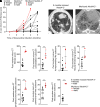
Micro-CT of end-stage IPF-like lung disease in moribund conditional Nedd4-2−/− mice. A: development of fibrosis scores in longitudinal micro-CT imaging studies of moribund conditional Nedd4-2−/− mice that had to be euthanized vs. conditional Nedd4-2−/− mice from the longitudinal study group that survived 5 mo of doxycycline induction. B: representative post mortem micro-CT images of a surviving conditional Nedd4-2−/− mouse and a moribund mouse that had to be euthanized after 4.5 mo. Summary of the post mortem micro-CT fibrosis scores (C), and the subscores for quantification of consolidation (D), honeycombing-like lesions (E), fissural thickening (F), peripheral bronchiectasis (G), parenchymal lines (H) and reticulations (I). n = 4–11 mice/group. *P < 0.05, **P < 0.01. IPF, idiopathic pulmonary fibrosis. micro-CT, microcomputed tomography.

References

    1. Raghu G, Remy-Jardin M, Richeldi L, Thomson CC, Inoue Y, Johkoh T, , et al. . Idiopathic pulmonary fibrosis (an update) and progressive pulmonary fibrosis in adults: an official ATS/ERS/JRS/ALAT clinical practice guideline. Am J Respir Crit Care Med 205: e18–e47, 2022. doi:10.1164/rccm.202202-0399ST. - DOI - PMC - PubMed
    1. King TE Jr, Pardo A, Selman M. Idiopathic pulmonary fibrosis. Lancet (Lancet 378: 1949–1961, 2011. doi:10.1016/S0140-6736(11)60052-4. - DOI - PubMed
    1. Martinez FJ, Collard HR, Pardo A, Raghu G, Richeldi L, Selman M, Swigris JJ, Taniguchi H, Wells AU. Idiopathic pulmonary fibrosis. Nat Rev Dis Primers 3: 17074, 2017. doi:10.1038/nrdp.2017.74. - DOI - PubMed
    1. Wolters PJ, Collard HR, Jones KD. Pathogenesis of Idiopathic Pulmonary Fibrosis. Annu Rev Pathol 9: 157–179, 2014. doi:10.1146/annurev-pathol-012513-104706. - DOI - PMC - PubMed
    1. Fernandez IE, Eickelberg O. New cellular and molecular mechanisms of lung injury and fibrosis in idiopathic pulmonary fibrosis. Lancet 380: 680–688, 2012. doi:10.1016/S0140-6736(12)61144-1. - DOI - PubMed

MeSH terms

Substances

LinkOut - more resources