

3T-3D FLAIR MRI in Menière's disease: associated profiles with clinical symptoms and electroacoustic characteristics
- PMID: 39438294
- PMCID: PMC11890404
- DOI: 10.1007/s00405-024-09029-6
3T-3D FLAIR MRI in Menière's disease: associated profiles with clinical symptoms and electroacoustic characteristics
Abstract
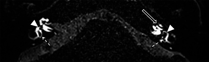
Purpose: Diagnosis of Menière's disease relies on clinical symptoms. Injected 3T MRI can show endolymphatic hydrops (EH), but correlation with the clinical status of MD, (probable -PMD or definite-DMD) remains doubtful. We revealed endolymphatic pressure disruption through functional exploration and verified if it was associated with an EH through MRI.
Materials and methods: We prospectively analyzed 3D3T FLAIR MRI of DMD and PMD patients. All of them underwent electrocochleography (EcoG), distortion-product otoacoustic emissions (DPOAEs), and videonystagmograhy (VNG). Amplitudes of summating potential (SP) and cochlear nerve action potential (AP) were measured on EcoG. DPOAE-phase was collected at 1 kHz for the 2f1-f2 DPOAE between sitting and laying position. A SP/AP ≥ 40% and a DPOAE phase-shift > 40° revealed pressure disruption.
Results: 39 patients (25 women, 53 y.o. 20-78), were included, with 32 DMD ears and 11 PMD ears. MRI was performed in a median of 21 days [0; 68] from the MD incident. Audiovestibular exploration took place 41 days after the crisis [0;83]. MRI revealed an EH in 71.9% and 27.2% of DMD and PMD, respectively. When combining functional explorations and MRI, testing was positive in 97% for DMD and 82% for PMD. When abnormal (59%), VNG mainly showed hyporeflexia in the diseased ear.
Conclusion: In patients suffering from DMD or PMD, with endolymphatic pressure disturbances confirmed by combined DPOAE-phase and EcoG, 3T 3D MRI reveals EH mostly in DMD but rarely in PMD. This seems to confirm that disturbance of endolymphatic pressure precedes EH.
Keywords: 3D3T-MRI; Distortion-product otoacoustic emissions; Electrocochleography; Hydrops; Meniére’s disease.
© 2024. The Author(s).
Figures


References
-
- Bruderer SG, Bodmer D, Stohler NA et al (2017) Population-based study on the epidemiology of Ménière’s Disease. Audiol Neurootol 22:74–82. 10.1159/000475875 - PubMed
-
- Huang D, Chen P, Chen S et al (2002) Expression patterns of aquaporins in the inner ear: evidence for concerted actions of multiple types of aquaporins to facilitate water transport in the cochlea. Hear Res 165:85–95. 10.1016/s0378-5955(02)00288-5 - PubMed
-
- Mhatre AN, Jero J, Chiappini I et al (2002) Aquaporin-2 expression in the mammalian cochlea and investigation of its role in Meniere’s disease. Hear Res 170:59–69. 10.1016/s0378-5955(02)00452-5 - PubMed
-
- Nakashima T, Pyykkö I, Arroll MA et al (2016) Meniere’s disease. Nat Rev Dis Primer 2:16028. 10.1038/nrdp.2016.28 - PubMed
MeSH terms
LinkOut - more resources
Full Text Sources
Medical
Miscellaneous
