. 2024 Oct 22;4(1):204.
doi: 10.1038/s43856-024-00623-7.

Advanced three-dimensional X-ray imaging unravels structural development of the human thymus compartments

Affiliations

Advanced three-dimensional X-ray imaging unravels structural development of the human thymus compartments

Savvas Savvidis et al. Commun Med (Lond). .

Abstract

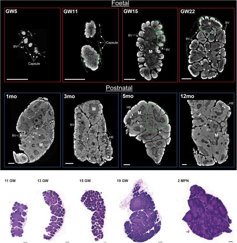
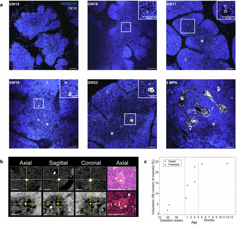
Background: The thymus, responsible for T cell-mediated adaptive immune system, has a structural and functional complexity that is not yet fully understood. Until now, thymic anatomy has been studied using histological thin sections or confocal microscopy 3D reconstruction, necessarily for limited volumes.

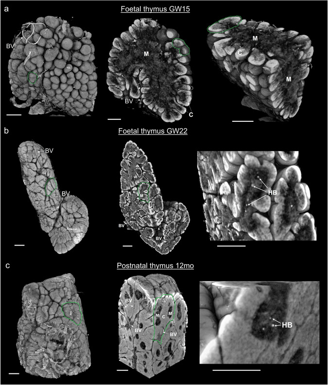
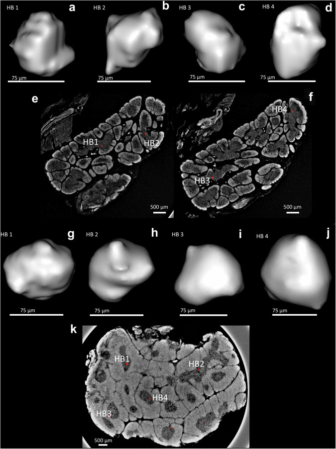
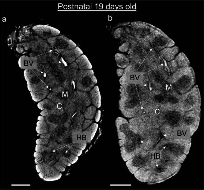
Methods: We used Phase Contrast X-Ray Computed Tomography to address the lack of whole-organ volumetric information on the microarchitecture of its structural components. We scanned 15 human thymi (9 foetal and 6 postnatal) with synchrotron radiation, and repeated scans using a conventional laboratory x-ray system. We used histology, immunofluorescence and flow cytometry to validate the x-ray findings.

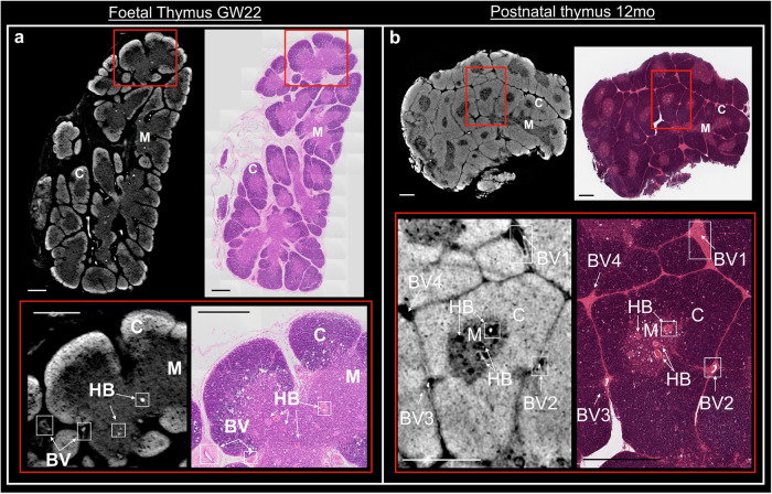
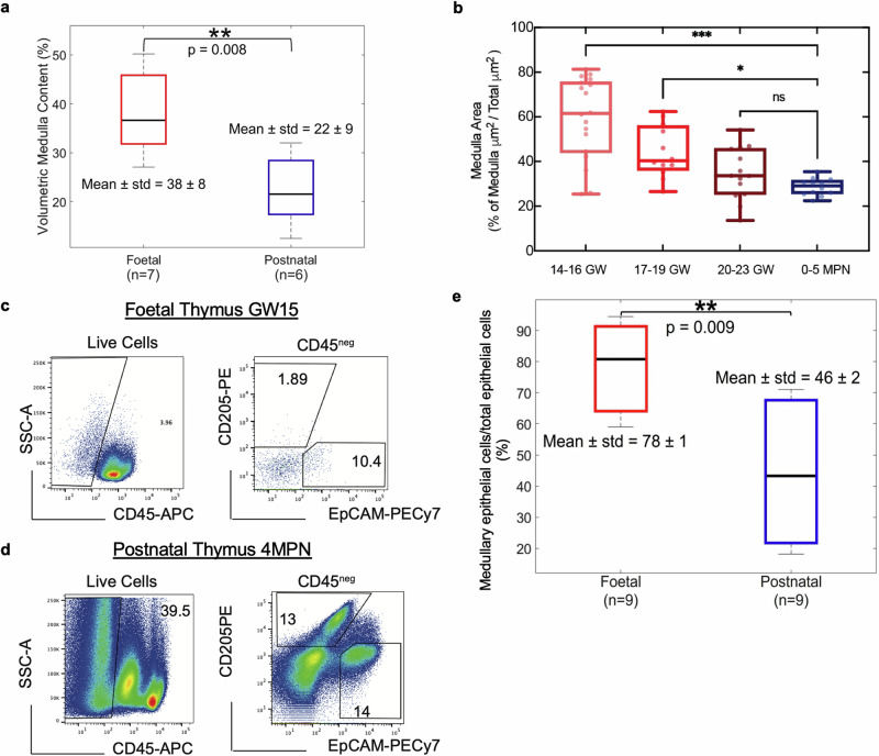
Results: Application to human thymi at pre- and post-natal stages allowed reliable tracking and quantification of the evolution of parameters such as size and distribution of Hassall's Bodies and medulla-to-cortex ratio, whose changes reflect adaptation of thymic activity. We show that Hassall's bodies can occupy 25% of the medulla volume, indicating they should be considered a third thymic compartment with possible implications on their role. Moreover, we demonstrate compatible results can be obtained with standard laboratory-based x-ray equipment, making this research tool accessible to a wider community.

Conclusions: Our study allows overcoming the resolution and/or volumetric limitations of existing approaches for the study of thymic disfunction in congenital and acquired disorders affecting the adaptive immune system.

Plain language summary

The thymus is the organ responsible for programming the immune system. It consists of two main compartments, named medulla and cortex. The medulla contains onion-shaped parts known as “Hassall’s bodies”. By imaging thymi at different stages of development with advanced x-ray methods, we gain understanding of changes that occur over time in 3D. We quantified how much of the thymus was occupied by these different components as they change with age, showing that Hassall’s bodies can take up 25% of the medulla, and should therefore be considered a proper part of the thymus with a purpose. Having a better understanding of the thymus can prove important in targeting conditions such as Down syndrome and thymic tumours, as well as provide information about structure.


Conflict of interest statement

The authors declare no competing interests.

Figures

Fig. 1
Fig. 1. Mapping of age associated structural changes of the thymic anatomical components via SPC-CT and histology.
a SPC-CT slices highlight the structural changes of the human thymus during maturation, starting from the foetal period to the early postnatal stages. The age of each sample in Carnegie Stage (CS), Gestational Week (GW) and month post-natal (MPN) is provided in the top left corner of each panel. Examples of areas undergoing lobulation (GW11 and GW15) and fully-formed lobules (GW22 and 5 MPN) are circled in green. The start of thymic corticomedullary definition is visible at GW11, and more evident at GW15, with compartmental organisation as a single central medulla and an outer cortex. Such conformation is repeated within each lobule from GW22 onwards. b Representative images of H&E staining on foetal and post-natal thymi supporting similar organogenesis as observed by SPC-CT. C cortex, M medulla, BVs blood vessels, HB Hassall bodies. Scale bars: 500 µm.
Fig. 2
Fig. 2. Intrasample comparison between SPC-CT and corresponding histological slices.
Reconstructed SPC-CT slices (left) and H&E staining (right) of a GW22 foetal thymus & b a 12 MPN thymus. The inset (red square) highlights an area of interest in each image, shown below, depicting typical anatomical aspects of the samples at their respective stage with exact correlation between the two imaging tools. C cortex, M medulla, BV blood vessel, HB Hassall bodies. Scale bars: 500 µm.
Fig. 3
Fig. 3. Volumetric visualisation and virtual dissection of the internal anatomy of the human thymus.
3D rendering of whole thymi (left images) and virtual dissection across two planes (centre & right images) at a. GW15, b GW22, and c 12MPN. Age-associated anatomical changes are evident from a to c; with lobule formation (indicated by green contours) starting from the external part of the organ (a), progressing throughout the organ (b) and eventually leading to a reduction in inter-lobular space (c). Corticomedullary organisation is observed as a single central medulla and outer cortex in foetal thymus GW15 (a), which is replicated in the individual lobules of foetal GW22 (b) and postnatal 12MPN (c). The morphology of the HBs is visualised volumetrically in b and c; a more detailed visualisation of their shape is provided in Fig. 6. C cortex, M medulla, HB Hassall’s body, BV Blood vessel. Scale bars: 500 µm.
Fig. 4
Fig. 4. Quantitative measures report age associated changes in thymic compartments.
a Volumetric quantification of medullary content in foetal (n = 7) and postnatal (n = 6) SPC-CT datasets. The foetal thymi demonstrated a significantly higher medulla content (38 ± 8%) than the postnatal thymi (22 ± 9%). 2 out of 9 foetal thymi (ages <GW11) were yet to start specialising into medulla and cortex compartments and were therefore excluded from this analysis. b Machine learning-based quantification of relative medullary area across development in H&E slices (n = 5 per group, with n = 3 technical triplicates). Samples were grouped into 4 categories, according to the developmental stage. Representative flow cytometry analysis of c foetal (GW15), and d postnatal (4 MPN) thymi. The percentage of EpCAMposCD205neg and CD205posEpCAMlow was calculated on total CD45 negative fraction, quantifying medullary and cortical epithelial cell percentages, respectively. The postnatal sample was negatively enriched for CD45 as previously described,. e Flow-cytometry based comparison of medullary epithelial cells content (EpCAMposCD205neg) relative to total epithelial cell counts. Results demonstrate a significantly higher medullary EC content in foetal thymi (n = 9, 78 ± 1) when compared to the post-natal counterparts (n = 9, 46 ± 2). p-values: <0.001(***); <0.005(**); >0.05(ns).
Fig. 5
Fig. 5. Qualitative and volumetric quantification of HBs across development.
a Representative immunofluorescence images (n = 10) showing appearance of KRT10 positive cells (grey) from GW16 with progressive development of HBs from GW19. Scale bars: 100 μm, nuclei counterstained with Hoescht. b Orthogonal views (axial, sagittal and coronal) of one of the HBs found in samples depicted in Fig. 2a and b, respectively. The corresponding histological slice (only axial plane available) is also shown. c SPC-CT based Volumetric HB content of the medulla as a function of developmental stage from foetal (n = 2) and postnatal (n = 6) thymi. Vertical lines subdivide the graph in three age ranges where the observed behaviour differs substantially. Scale bars: 100 μm.
Fig. 6
Fig. 6. Individually segmented Hassall’s bodies.
ad Show four separately segmented HBs alongside their original position in SPC-CT slices of a foetal thymus (e, f); gj show the same for the postnatal case, in which a single SPC-CT slice (k) was sufficient thanks to the increased HB presence. In both cases, a mild smoothing based on mesh interpolation was applied to avoid an excessively “blocky” appearance caused by the limited number of voxels an HB consists of.
Fig. 7
Fig. 7. Laboratory-based CT solutions provide the same structural information as synchrotron equipment.
Corresponding slices of a 19-day post-natal thymus acquired using SPC-CT (a) and H-CT (b). Both depict the same structural components of this sample, including cortex (C), medulla (M), Hassall’s bodies (HB) and blood vessels (BV). Scale bars: 500 µm.

References

    1. Asghar, A., Asad, M. R., Naaz, S. & Rani, M. Screening of the growth of thymus of human fetuses. Anat. Cell Biol.52, 478–485 (2019). - PMC - PubMed
    1. Mizuki Nishino, S. K. A., Kocher, O. N., Robert, L., Thurer, P. M. B. & Hatabu, H. The Thymus: A Comprehensive Review. Radiogr2006, 335–348 (2006). - PubMed
    1. Farley, A. M. et al. Dynamics of thymus organogenesis and colonization in early human development. Dev140, 2015–2026 (2013). - PMC - PubMed
    1. Gayathri, J., Bharathadevi, M. & Sivakami, T. a Study on Histogenesis of Thymus in Human Foetuses. Int. J. Anat. Res.7, 6811–6817 (2019).
    1. Varga, I., Pospisilova, V., Jablonska-Mestanova, V., Galfiova, P. & Polak, S. The thymus: picture review of human thymus (renatal development). Bratisl. Lek. Listy112, 368–376 (2011). - PubMed

LinkOut - more resources