

Spatial attention-based CSR-Unet framework for subdural and epidural hemorrhage segmentation and classification using CT images
- PMID: 39438833
- PMCID: PMC11494839
- DOI: 10.1186/s12880-024-01455-6
Spatial attention-based CSR-Unet framework for subdural and epidural hemorrhage segmentation and classification using CT images
Abstract
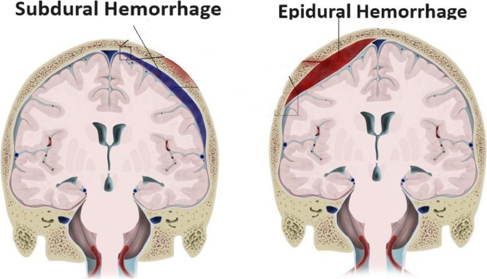
Background: Automatic diagnosis and brain hemorrhage segmentation in Computed Tomography (CT) may be helpful in assisting the neurosurgeon in developing treatment plans that improve the patient's chances of survival. Because medical segmentation of images is important and performing operations manually is challenging, many automated algorithms have been developed for this purpose, primarily focusing on certain image modalities. Whenever a blood vessel bursts, a dangerous medical condition known as intracranial hemorrhage (ICH) occurs. For best results, quick action is required. That being said, identifying subdural (SDH) and epidural haemorrhages (EDH) is a difficult task in this field and calls for a new, more precise detection method.
Methods: This work uses a head CT scan to detect cerebral bleeding and distinguish between two types of dural hemorrhages using deep learning techniques. This paper proposes a rich segmentation approach to segment both SDH and EDH by enhancing segmentation efficiency with a better feature extraction procedure. This method incorporates Spatial attention- based CSR (convolution-SE-residual) Unet, for rich segmentation and precise feature extraction.
Results: According to the study's findings, the CSR based Spatial network performs better than the other models, exhibiting impressive metrics for all assessed parameters with a mean dice coefficient of 0.970 and mean IoU of 0.718, while EDH and SDH dice scores are 0.983 and 0.969 respectively.
Conclusions: The CSR Spatial network experiment results show that it can perform well regarding dice coefficient. Furthermore, Spatial Unet based on CSR may effectively model the complicated in segmentations and rich feature extraction and improve the representation learning compared to alternative deep learning techniques, of illness and medical treatment, to enhance the meticulousness in predicting the fatality.
Keywords: Classification; Deep Learning; Epidural hemorrhage; Intracranial hemorrhage; Segmentation; Subdural hemorrhage.
© 2024. The Author(s).
Conflict of interest statement
The authors declare no competing interests.
Figures


References
-
- Xi G, Keep RF, Hoff JT. Mechanisms of brain injury after intracerebral haemorrhage. Lancet Neurol. 2006;5(1):53–63. - PubMed
-
- Foreman KJ, Marquez N, Dolgert A, Fukutaki K, Fullman N, McGaughey M, Pletcher MA, Smith AE, Tang K, Yuan CW, Brown JC. Forecasting life expectancy, years of life lost, and all-cause and cause-specific mortality for 250 causes of death: reference and alternative scenarios for 2016–40 for 195 countries and territories. Lancet. 2018;392(10159):2052–90. - PMC - PubMed
-
- Chang CS, Chang TS, Yan JL, Ko L. All attention U-NET for semantic segmentation of intracranial hemorrhages in head CT images. In: 2022 IEEE Biomedical Circuits and Systems Conference (BioCAS). USA: IEEE; 2022. p. 600–4.
-
- Skalski M. Diagram - intracranial hemorrhage. Case study, Radiopaedia.org. 10.53347/rID-21542. Accessed 25 May 2024.
MeSH terms
LinkOut - more resources
Full Text Sources
Medical
