

Is the identification of caseating granuloma in the intestine indicative of tuberculosis? a rare case of Crohn's disease
- PMID: 39438897
- PMCID: PMC11494966
- DOI: 10.1186/s13000-024-01566-2
Is the identification of caseating granuloma in the intestine indicative of tuberculosis? a rare case of Crohn's disease
Abstract
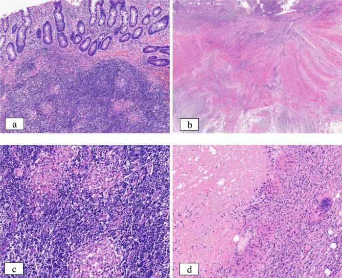
Background: Crohn's disease (CD) is a chronic intestinal inflammatory disorder, the etiology of which remains unknown, and is characterized by symptoms such as chronic abdominal pain, diarrhea, obstruction, and perianal lesions. Histopathology is widely regarded as the preferred method for diagnosing CD, although the histological diagnosis may lack specificity. The identification of granulomas is commonly believed to be the most reliable diagnostic indicator for CD, surpassing all other clinical features in significance. Nevertheless, research indicates that the detection rate of granulomas in CD exhibits considerable variability. Furthermore, granulomas can manifest in various specific infections including tuberculosis and Yersinia, as well as in a range of diseases characterized by macrophage reactions such as sarcoidosis and drug-induced enteritis. Granulomas associated with CD typically do not exhibit necrosis. However, the formation of caseous granulomas may occur as a result of secondary infections related to anti-CD drug treatment or perforation of the intestinal wall.
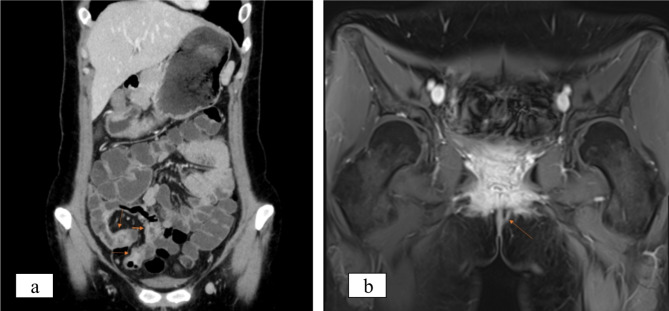
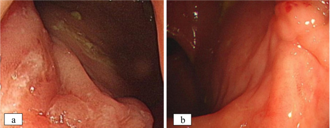
Case presentation: In this study, we present a case of a 28-year-old female patient diagnosed with CD exhibiting histologic granulomas, including both caseating and non-caseating forms, which demonstrated a positive response to medical treatment.
Conclusion: In clinical practice, various forms of granulomas may indicate diverse underlying diseases, yet lack specificity. It is suggested that the presence of caseous granulomas should not be considered as a definitive exclusion criterion for the diagnosis when clinical, endoscopic, imaging and other histopathological features are consistent with CD. This study is the first report of caseous granulomas in CD without concomitant tuberculosis infection.
Keywords: Case report; Caseating granuloma; Crohn’s disease; Specific bacterial infections; Tuberculosis.
© 2024. The Author(s).
Conflict of interest statement
The authors declare no competing interests.
Figures
Similar articles
-
Significance of granuloma and granulomatous lymphangitis in the differential diagnosis of Crohn's disease.J Dig Dis. 2020 Aug;21(8):454-461. doi: 10.1111/1751-2980.12919. J Dig Dis. 2020. PMID: 32621394
-
CD73 expression in tissue granulomas in distinguishing intestinal tuberculosis from Crohn's disease in a South African cohort.Scand J Gastroenterol. 2018 Oct-Nov;53(10-11):1217-1221. doi: 10.1080/00365521.2018.1503326. Epub 2018 Oct 8. Scand J Gastroenterol. 2018. PMID: 30295112
-
[Granulomas in Intestinal Biopsies? Intestinal Tuberculosis Mimicking Crohn's Disease: A Case Report].Rev Med Chil. 2024 Jun;152(6):730-735. doi: 10.4067/s0034-98872024000600730. Rev Med Chil. 2024. PMID: 39760568 Spanish.
-
Differentiating Crohn's disease from intestinal tuberculosis.World J Gastroenterol. 2019 Jan 28;25(4):418-432. doi: 10.3748/wjg.v25.i4.418. World J Gastroenterol. 2019. PMID: 30700939 Free PMC article. Review.
-
Clinical significance of granulomas in Crohn's disease: A systematic review and meta-analysis.J Gastroenterol Hepatol. 2020 Mar;35(3):364-373. doi: 10.1111/jgh.14849. Epub 2019 Nov 26. J Gastroenterol Hepatol. 2020. PMID: 31522456
References
-
- Hessian PA, Highton J, Kean A, Sun CK, Chin M. Cytokine profile of the rheumatoid nodule suggests that it is a Th1 granuloma. Arthritis Rheum. 2003;48:334–8. - PubMed
-
- Brown I, Kumarasinghe MP. Granulomas in the gastrointestinal tract: deciphering the Pandora’s box. Virchows Arch. 2018;472:3–14. - PubMed
-
- Torres J, Mehandru S, Colombel JF, Peyrin-Biroulet L. Crohn’s disease. Lancet. 2017;389:1741–55. - PubMed
-
- Leong RWL. The significance of granulomas in Crohn’s disease and inflammatory bowel disease epidemiology in Asia. J Gastroenterol Hepatol. 2020;35:523–24. - PubMed
Publication types
MeSH terms
LinkOut - more resources
Full Text Sources
Medical
