Review
. 2024 Oct 22;14(1):130.
doi: 10.1186/s13578-024-01311-8.

Golgiphagy: a novel selective autophagy to the fore

Affiliations
Review

Golgiphagy: a novel selective autophagy to the fore

Yifei Chen et al. Cell Biosci. .

Abstract

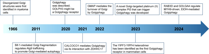
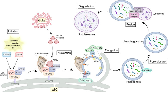
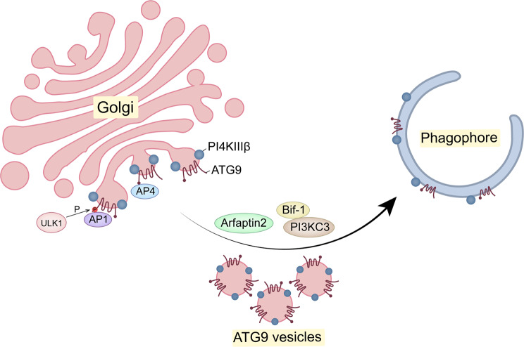
The Golgi apparatus is the central hub of the cellular endocrine pathway and plays a crucial role in processing, transporting, and sorting proteins and lipids. Simultaneously, it is a highly dynamic organelle susceptible to degradation or fragmentation under various physiological or pathological conditions, potentially contributing to the development of numerous human diseases. Autophagy serves as a vital pathway for eukaryotes to manage intracellular and extracellular stress and maintain homeostasis by targeting damaged or redundant organelles for removal. Recent research has revealed that autophagy mechanisms can specifically degrade Golgi components, known as Golgiphagy. This review summarizes recent findings on Golgiphagy while also addressing unanswered questions regarding its mechanisms and regulation, aiming to advance our understanding of the role of Golgiphagy in human disease.

Keywords: Autophagy; Golgi apparatus; Golgi fragmentation; Golgiphagy; Receptor.


Conflict of interest statement

The authors declare that they have no competing interests.

Figures

Fig. 1
Fig. 1
The key discoveries in the Golgiphagy field
Fig. 2
Fig. 2
Core machinery of autophagy. In response to various stress conditions, the mTORC1 and AMPK pathways regulate the kinase activity of the ULK1 complex, thereby initiating autophagy. The activated ULK1 complex is recruited near the ER membrane due to the interaction of FIP200 with VAPA/VAPB on the ER. Additionally, ATG9 vesicles are recruited to the ER membrane to provide a membrane source through interaction with the ATG13-ATG101 subcomplex. Subsequently, ULK1 further activates the PI3KC3 complex 1, resulting in the generation of PI3P. WIPI2 then binds to PI3P and further recruits the ATG12-ATG5-ATG16L1 complex to phagophore, thereby mediating ATG8 lipidation. Concurrently, PI3P also recruits WIPI3 or WIPI4 to phagophore, where WIPI3/4 transfer phospholipids from the ER to phagophore through interactions with ATG2, ATG9, as well as VMP1 and TMEM41B on the ER, thereby promoting phagophore expansion. Subsequently, the autophagosome closes through the action of the ESCRT mechanism, after which specific SNARE proteins, HOPS complexes, and small GTPases such as RAB7 mediate the fusion of the autophagosome with the lysosome. Following fusion, the cargo is degraded by hydrolytic enzymes within the lysosome and reused by the cell. Abbreviations: mTORC1, mammalian target of rapamycin complex 1; AMPK, adenosine 5’-monophosphate-activated protein kinase; ULK1, UNC51-like kinase; ER, endoplasmic reticulum; FIP200, focal adhesion kinase family interacting protein of 200 kD; VAPA, VAMP (Vesicle Associated Membrane Protein)-associated protein A; VAPB, VAMP (Vesicle Associated Membrane Protein)-associated protein B; ATG, autophagy-related; PI3KC3, phosphoinositide 3-kinases catalytic subunit type 3; PI3P, phosphatidylinositol 3-phosphate; WIPI, WD repeat structural domain phosphatidylinositol-interacting protein; VMP1, Vacuole membrane protein 1; TMEM41B, Transmembrane Protein 41B; ESCRT, endosomal sorting complex required for transport; SNARE, soluble N-ethylmaleimide-sensitive factor attachment protein receptors; HOPS, homotypic fusion and vacuole protein sorting; RAB, Ras analog in brain
Fig. 3
Fig. 3
The involvement of Golgi in ATG9 trafficking during autophagy. ATG9 vesicles are transported from the Golgi compartment to autophagosome formation sites (phagophore) via AP1 and/or AP4 complexes, which also deliver PI4KIIIβ. Furthermore, ULK1 phosphorylates ATG9, thereby facilitating ATG9 binding to AP1 complexes. The correct delivery of ATG9 vesicles to phagophore is also regulated by Arfaptin2, Bif-1, and the PI3KC3 complex. Abbreviations: ATG, autophagy-related; AP, adaptor protein; PI4KIIIβ, phosphatidylinositol-4-kinase-IIIβ; ULK1, UNC51-like kinase; Bif-1, BAX-interacting protein 1; PI3KC3, phosphoinositide 3-kinases catalytic subunit type 3
Fig. 4
Fig. 4
The structure of the Golgiphagy receptors. (A) Schematic structure of GOLPH3 protein. GPP34, PI4P-binding domain. (B) Schematic structure of CALCOCO1 protein. SKICH, SKIP carboxyl homology domain; LIR, LC3-interacting region; CC, coil-coil region; zDABM, zDHHC-AR-binding motif; UIR, UDS-interacting region; ZF, Zinc Finger; FFAT, two phenylalanines in an acidic tract domain. (C) Schematic structure of dGMAP protein. GRAB, GRIP-related Arf-binding domain. (D) Schematic structures of the YIPF3 protein and the YIPF4 protein. TMD, transmembrane domain. (E) A table of the amino acid sequences as well as the positions of the LIR motifs in these Golgiphagy receptors
Fig. 5
Fig. 5
Models of Golgiphagy receptors-mediated Golgiphagy upon nutrient starvation in mammalian or Drosophila melanogaster. (A) GOLPH3, which is located on the trans-Golgi by binding PI4P, interacts with ATG8 to promote the encapsulation of Golgi fragments by phagophore. (B) CALCOCO1 localizes to the Golgi apparatus through its zDABM motif binding to the AR domain of ZDHHC17. Subsequently, CALCOCO1 interacts with ATG8 through its LIR and UIR motifs, recruiting the autophagy machinery and thus facilitating the degradation of Golgi fragments. (C) In Drosophila melanogaster, dGMAP can directly bind to ATG8 through the N-terminal LIR motif, thereby mediating the autophagy pathway degradation of the Golgi fragments. (D) YIPF3 and YIPF4, which are anchored to the Golgi through 5 tightly stacked transmembrane domains, form a heterodimer. As the only membrane-embedded Golgiphagy receptors, they interact with ATG8 to mediate Golgiphagy. Abbreviations: GOLPH3, Golgi phosphoprotein 3; CALCOCO1, calcium binding and coiled-coil domain protein 1; dGMAP, Golgi microtubule-associated protein in Drosophila melanogaster; YIPF3, the member 3 of Yip1 domain family; YIPF4, the member 4 of Yip1 domain family; PI4P, phosphatidylinositol 4-phosphate; AR, ankyrin repeat; SKICH, SKIP carboxyl homology; LIR, LC3-interacting region; CC, coil‐coil regions; zDABM, zDHHC-AR-binding motif; UIR, UDS‐interacting region; ZF, Zinc Finger; FFAT, two phenylalanines in an acidic tract
Fig. 6
Fig. 6
The process and regulation of Golgiphagy. Under various external stimuli, including starvation, alcohol exposure and other extreme situations, the Golgi apparatus undergoes fragmentation, which in turn triggers the process of Golgiphagy. The Golgiphagy receptors, localized on the Golgi, mediate phagophore wrapping around the fragmented Golgi by binding to ATG8 on the phagophore. The phagophore continue to extend and then close, forming autophagosomes which in turn fuse with lysosomes thereby degrading Golgi components. The process of Golgiphagy is subject to regulation by several factors. (A) Bif-1 could rupture the Golgi membrane during starvation, thereby inducing Golgiphagy. (B) RAB3D, MYH10, and GOLGA4 form a complex to maintain the structural integrity of the Golgi apparatus. Upon ethanol treatment, RAB3D is reduced, MYH10 separates from the Golgi, and MYH9 exerts force to disperse the Golgi membrane by binding to it via RAB6A. Concurrently, the conformation of the GOLGA4 protein is altered to facilitate the formation of the phagophore from the fragmented Golgi cisterna. (C) In conditions of starvation, WAC inhibits the binding of GABARAP to GM130, thereby allowing the maintenance of the centrosomal GABARAP pool. The centrosomal GABARAP is transported along microtubules to the phagophore, where it mediates the autophagic activation of the ULK1 complex and promotes the biogenesis of autophagosomes during Golgiphagy. Abbreviations: ATG, autophagy-related; Bif-1, BAX-interacting protein 1; RAB, Ras analog in brain; MYH10/NMIIB, non-muscle myosin II B; MYH9/NMIIA, non-muscle myosin II A; GOLGA4, golgin A4; WAC, WW domain-containing adaptor with coiled coil; GM130/GOLGA2, Golgin subfamily A member 2; GABARAP, GABA Type A Receptor-Associated Protein; ULK1, UNC51-like kinase

References

    1. Bentivoglio M. 1898: the golgi apparatus emerges from nerve cells. Trends Neurosci. 1998;21:195–200. - PubMed
    1. Short B, Barr FA. The golgi apparatus. Curr Biol CB. 2000;10:R583–585. - PubMed
    1. Potelle S, Klein A, Foulquier F. Golgi post-translational modifications and associated diseases. J Inherit Metab Dis. 2015;38:741–51. - PubMed
    1. De Matteis MA, Luini A. Exiting the golgi complex. Nat Rev Mol Cell Biol. 2008;9:273–84. - PubMed
    1. Wei J-H, Seemann J. Unraveling the golgi ribbon. Traffic Cph Den. 2010;11:1391–400. - PMC - PubMed

LinkOut - more resources