. 2024 Dec;11(45):e2405711.
doi: 10.1002/advs.202405711. Epub 2024 Oct 22.

A Tympanic Piezo-Bioreactor Modulates Ion Channel-Associated Mechanosignaling to Stabilize Phenotype and Promote Tenogenesis in Human Tendon-Derived Cells

Affiliations

A Tympanic Piezo-Bioreactor Modulates Ion Channel-Associated Mechanosignaling to Stabilize Phenotype and Promote Tenogenesis in Human Tendon-Derived Cells

Marc A Fernandez-Yague et al. Adv Sci (Weinh). 2024 Dec.

Abstract

Preserving the function of human tendon-derived cells (hTDCs) during cell expansion is a significant challenge in regenerative medicine. In this study, a non-genetic approach is introduced to control the differentiation of hTDCs using a newly developed tympanic bioreactor. The system mimics the functionality of the human tympanic membrane, employing a piezoelectrically tuned acoustic diaphragm made of polyvinylidene fluoride-co-trifluoroethylene and boron nitride nanotubes. The diaphragm is vibrationally actuated to deliver targeted electromechanical stimulation to hTDCs. The results demonstrate that the system effectively maintains the tendon-specific phenotype of hTDCs, even under conditions that typically induce nonspecific differentiation, such as osteogenesis. This stabilization is achieved by modulating integrin-mediated mechanosignaling via ion channel-regulated calcium activity, potentially by TREK-1 and PIEZO1, yet targeted studies are required for confirmation. Finally, the system sustains the activation of key differentiation pathways (bone morphogenetic protein, BMP) while downregulating osteogenesis-associated (mitogen-ctivated protein kinase, MAPK and wingless integrated, WNT) pathways and upregulating Focal Adhesion Kinase (FAK) signaling. This approach offers a finely tunable, dose-dependent control over hTDC differentiation, presenting significant potential for non-genetic approaches in cell therapy, tendon tissue engineering, and the regeneration of other mechanosensitive tissues.

Keywords: BNNT; FAK; PVDF‐TrFE; electromechanical; focal adhesions; mechanotransduction; piezoelectricity; tendon.


Conflict of interest statement

The authors declare no conflict of interest.

Figures

Figure 1
Figure 1
Fabrication of a Piezoelectrically Tunable PVDF‐TrFE Nanocomposite Membrane. a) Schematic illustrations of steps in fabricating PVDF‐TrFE/BNNT nanocomposite films and EMS using spin‐coating. A PVDF‐TrFE polymeric solution is applied onto a glass substrate, rapidly spun, and air‐dried to create a uniform thin film. The combination of centripetal forces and surface tension ensures a homogenous coating. b) The production of thin films with a 5–10 µm thickness after 30 s at 1000 RPMs is depicted, where the process is repeated three times to obtain films with an average thickness of 25 ± 5 µm. c) TEM image of aggregated BNNTs attributed to the polarity of the B─N bond. d) The Hansen parameters are graphically represented for different solvent and co‐solvent systems, denoted by coordinates of dispersion (δd), polar (δp), and hydrogen (δh) bonding. The sphere illustrates the Hansen parameters of BNNTs, which achieved a stable dispersion in the solution for over 24 h. e) Increased polar crystallinity, as observed in electrically poled and thermally annealed films at 132 °C for 2 h, is indicated by XRD analysis. f) Depiction of the d33 coefficient increases as a function of the poling field in PVDF‐TrFE/BNNT films. g) XRD analyses of pristine and nanocomposite samples before and after poling. h) XRD curves illustrate the quantification of polar and non‐polar phase fractions. i) Representative FTIR spectra showing the peaks and associated phase identification (n = 3, r = 2). j) Representative out‐of‐plane hysteresis loops demonstrating successful incorporation of BNNTs and piezoelectric behavior (n = 3, r = 6). P.T. refers to PVDF‐TrFE. Unpoled samples showed no hysteresis or piezoresponse.
Figure 2
Figure 2
Piezobioreactor Multi‐Resonant Configuration for Tunable Piezoelectric Performance and Frequency Selectivity of Mechanically Actuated Acoustic Diaphragm. Schematic representation of tympanic membrane function a): Vibration‐wave, depicted as alternating high‐and low‐pressure waves, impact the tympanic membrane, causing it to resonate. This vibration transfers energy through the middle ear as mechanical amplifications, represented by the gain transfer function graph. The inset shows the oscillation map of the tympanic membrane under stimulation. b) Schematic representation of the piezobioreactor diaphragm configuration, with the membrane tension precisely modulated for an optimal vibrational response. COMSOL modeling output showcasing the harmonic response of the membrane within the frequency range of 0–2000 Hz at a set actuator amplitude of 30 nm. Below ≈700 Hz, the membrane response remains in phase with the input vibration, as detailed in the magnified inset (100–200 Hz). Beyond this frequency, resonant modes appear (≈700 Hz, ≈1500 Hz, and ≈2500 Hz), leading to significant amplification and deformation of the membrane, as depicted by the model's minima and maxima amplitude data. c) Sample electrical responses as captured by voltameters with two distinct inner impedances (70 MΩ and 200 GΩ) (top) juxtaposed against the corresponding input signal (bottom). d) Voltage output as a frequency sweep function for unpoled pristine, poled pristine, and poled composite PVDF‐TrFE films. e) Total displacement and electric potential as a function of frequency sweep, obtained through scanning laser interferometry‐based voltammetry; the data points represent actual measurements, while the curves represent COMSOL modeling outputs (n = 7 samples).
Figure 3
Figure 3
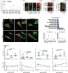
Mechanical stimulation drives osteogenic pathways while inhibiting tenogenic differentiation in MSCs. Nanoscale vibrational stimulation (MS) led to a significant downregulation in the expression of a) TNMD and, b) SCX indicating a deviation from tenogenic lineage, using tendon cells as a reference for baseline expression levels. Simultaneously, an upregulation in the expression of c) BMP and, d) WNT was observed, suggesting the onset of osteogenic differentiation. e) FAK indicates an increase in cell adhesion signaling as a response to MS. f) Ion channels expression changes response to MS at days 1 and 5. Data is represented as Median ± range (N = 4, r = 6). Statistical analysis was performed using Non‐parametric Kruskal–Wallis followed by Dunn's multiple‐comparisons test. Statistical significance is indicated as follows: * p < 0.05, ** p < 0.01, *** p < 0.005, **** p < 0.001.
Figure 4
Figure 4
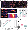
Mechanical Stimulation Modulates Ca2+ Events in hTDCs. a) Fluo‐4 imaging illustrates Ca2+ signals at baseline (Control, left), and during MS (middle) and EMS (right) both at 1500 Hz. Insets provide magnified views, emphasizing changes in Ca2+ intensity. Scale bars: 100 µm (inset 20 µm). b) Time‐lapse imaging (left) depicts Ca2+ waves originating from the cell periphery (arrows). Scale bars: 20 µm. The right panel shows a kymograph of a representative cell. c) Representative Ca2+ signals for Control (top), MS (middle), and EMS (bottom). d) Fraction of cells responding to stimuli (n = 112 cells for Control, n = 99 for MS, and n = 136 for EMS from 3 independent experiments). Tukey's multiple‐comparison test: p < 0.001. One‐way ANOVA test was used: * p < 0.05, ** p < 0.01 and NS. Full statistics are in the Methods. g) Cumulative distribution of response amplitude (ΔF/F0) for the different stimulus. The 75th percentile ΔF/F 0 values for each group: 1.04 (Control), 1.11 (EMS), 1.29 (MS) (n = 112 cells for Control, n = 99 for MS, and n = 136 for EMS from 3 independent experiments). Full statistics are in the Methods.
Figure 5
Figure 5
EMS Modulates hTDCs Functional Response. a) Evaluation of protein expression levels related to tendon maturation and collagen synthesis at days 1 and 5 in hTDCs cultured under MS and EMS at 1500 Hz. Data are presented as Median ± IQR (N = 4). b) Effect of inhibiting ROCK/RhoA (Y27623) or Myosin II activity (blebbistatin) on tendon‐specific markers expression, (N = 4). c) Effect of inhibiting ion channels activity (Gd3+) on collagens, integrin receptors, and d) on ion channels expression (N = 4). e) Paxillin expression as a marker for cell adhesion signaling (N = 3). f) Gene ontology pathway analysis showing the activation of specific biological pathways induced by EMS (top). Gene expression fold‐change (bottom) in cells subjected to EMS (D5, N = 3). g) Protein expression (top) and correlative analysis (bottom) for TGF‐β, h) WNT i) MAPK, and j) FAK activation under different EMS levels (0, 1, and 3 mV). Description of pathway activation is in the Experimental Section. Data is represented as Median ± range (N = 3, with the experiment replicated six times). Statistical analysis was performed using Non‐parametric Kruskal–Wallis followed by Dunn's multiple‐comparisons test. The representation of statistically significant differences is denoted as * p < 0.05, ** p < 0.01, *** p < 0.005, **** p < 0.001.

References

    1. Bi Y., Ehirchiou D., Kilts T. M., Inkson C. A., Embree M. C., Sonoyama W., Li Li, Leet A. I., Seo B.‐M., Zhang Li, Shi S., Young M. F., Nat. Med. 2007, 13, 1219. - PubMed
    1. Zhang J., Wang J. H., Methods Mol. Biol. 2018, 1842, 217. - PubMed
    1. Wei B., Lu J., Stem Cell Rev. Rep. 2021, 17, 1534. - PubMed
    1. Nature 2023, 616, 629. - PubMed
    1. Lomas A. J., Ryan C. N. M., Sorushanova A., Shologu N., Sideri A. I., Tsioli V., Fthenakis G. C., Tzora A., Skoufos I., Quinlan L. R., O'Laighin G., Mullen A. M., Kelly J. L., Kearns S., Biggs M., Pandit A., Zeugolis D. I., Adv. Drug Deliv. Rev. 2015, 84, 257. - PubMed

LinkOut - more resources