. 2024 Oct 7;15(18):6122-6134.
doi: 10.7150/jca.100651. eCollection 2024.

Development and Validation of a Nomogram for Axillary Lymph Node Metastasis Risk in Breast Cancer

Affiliations

Development and Validation of a Nomogram for Axillary Lymph Node Metastasis Risk in Breast Cancer

Shijing Wang et al. J Cancer. .

Abstract

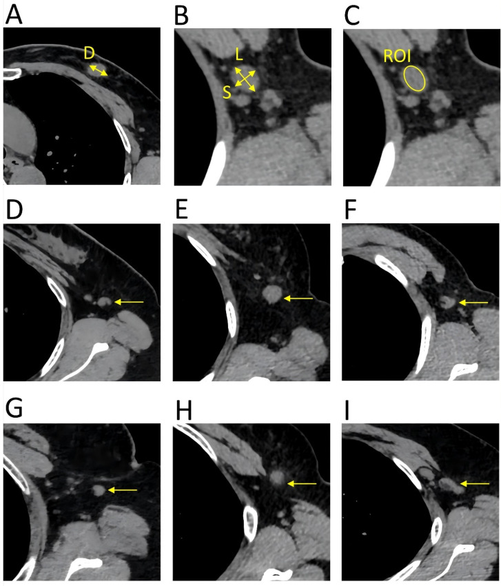
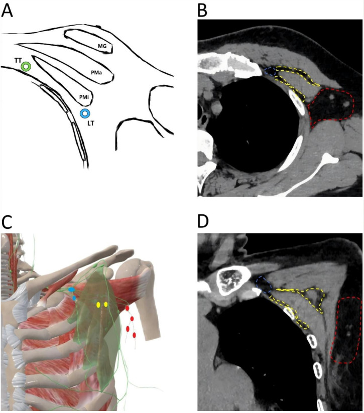
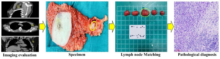
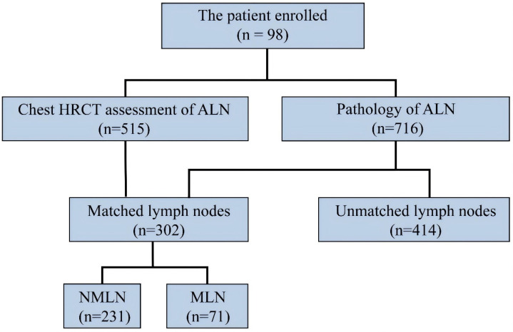
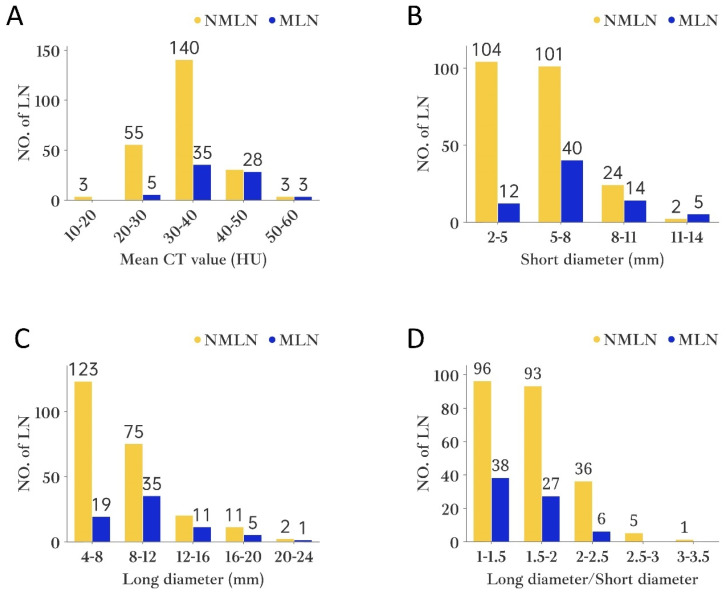
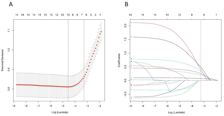
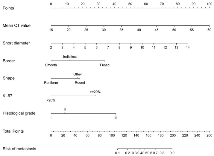
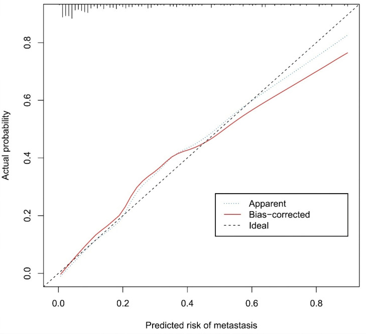
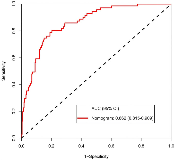
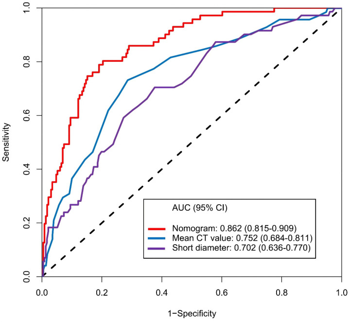
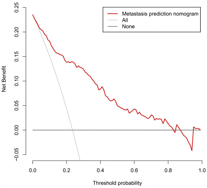
Purpose: Preoperative assessment of axillary lymph node (ALN) status is essential for breast cancer treatment planning. This study prospectively analyzed risk factors for ALN metastasis by comparing high-resolution computed tomography (HRCT) imaging with pathology and developed a nomogram to aid in diagnosis. Methods: From April 2023 to May 2024, breast cancer patients confirmed by pathology participated in the study. All had chest HRCT before surgery, and ALN samples were anatomically matched to HRCT imaging and pathology. The least absolute shrinkage and selection operator (LASSO) regression helped refine metastasis features, and a nomogram was constructed using the final selected features determined by multivariate logistic regression. The nomogram's performance was evaluated with concordance index (C-index), calibration plot, and decision curve analysis, with internal validation through bootstrapping. Results: A total of 302 ALN from 98 patients were included in this study. The predictors included in the nomogram encompassed the mean CT value, short diameter, border, and shape of ALN, as well as the Ki-67 status and histological grade of the primary tumor. The model exhibited satisfactory discrimination, with a C-index of 0.869 (95% CI: 0.826-0.912) and an AUC of 0.862 (95% CI, 0.815-0.909). The calibration curve demonstrated a high degree of concordance between the predicted and actual probabilities. The decision curve analysis demonstrated that the nomogram was clinically useful when the threshold for intervention was set at the metastasis possibility range of 1% to 86%. Conclusion: The nomogram combined with preoperative pathology and HRCT imaging have the potential to improve the evaluation of ALN status.

Keywords: Axillary lymph node; Breast cancer; HRCT; Metastasis; Nomogram.


Conflict of interest statement

Competing Interests: The authors have declared that no competing interest exists.

Figures

Figure 1
Figure 1
Key parameters of the HRCT imaging. The primary tumor and lymph nodes were evaluated in the transverse of the HRCT imaging, with a slice thickness of 1 mm. A. (D) diameter of the primary tumor. B. (L) long diameter of the lymph node; (S) short diameter of the lymph node. C. The mean CT value was calculated by delineating a region of interest that encompassed the entire lymph node, ensuring that the measurement line remained within the border of the lymph node. D. Reniform shape. E. Round shape. F. Other shape. G. Smooth border. H. Indistinct border. I. Fused border.
Figure 2
Figure 2
Anatomical location of axillary lymph node. A. Transverse anatomical schematic. MG: mammary gland, Pma: pectralis major muscle, Pmi: petcoralis minor muscle, LT: lateral thoracic artery (blue), TT: thoracic acromial artery (green). B. Anatomical location of axillary lymph node in a CT transverse view. The area where the level I axillary lymph node is located (red); The area where the level II axillary lymph node is located (yellow); The area where the level III axillary lymph node is located (blue). C. Coronal anatomical schematic. The pectoralis minor muscle (light green), level I axillary lymph node (red), level II axillary lymph node (yellow), level III axillary lymph node (blue). D. Anatomical location of axillary lymph node in a CT coronal view. The area where the level I axillary lymph node is located (red); The area where the level II axillary lymph node is located (yellow); The area where the level III axillary lymph node is located (blue).
Figure 3
Figure 3
Flow chart comparing imaging to pathology. CT detected a 2x1.5cm lymph node located laterally to the left pectoralis minor. The node was surgically located, and subsequent pathology confirmed breast cancer metastasis.
Figure 4
Figure 4
Imaging versus the pathology in 716 lymph node of 98 patients. Abbreviations: ALN-axillary lymph node; MLN-metastasis lymph node; NMLN-non-metastasis lymph node.
Figure 5
Figure 5
The histogram of axillary lymph node distribution under different variables. A. Mean CT value; B. Short diameter; C. Long diameter; D. Long diameter/Short diameter. Abbreviations: NO.of LN- number of lymph nodes.
Figure 6
Figure 6
Selection of HRCT imaging and pathology features using the LASSO regression. A. Optimal lambda selection in the LASSO model involved fivefold cross-validation based on minimum criteria. The binomial deviance curve was plotted against log(lambda), with dotted lines indicating optimal values determined by both the minimum criteria and the 1-SE criteria (red). B. The LASSO coefficient profiles for 14 features were plotted versus log (lambda). A vertical line (red) indicates the optimal lambda chosen through fivefold cross-validation, resulting in six features with nonzero coefficients. Abbreviations: HRCT-high-resolution computed tomography; LASSO-least absolute shrinkage and selection operator; SE-standard error.
Figure 7
Figure 7
Developed prediction nomogram. The axillary lymph node metastasis nomogram was developed in the cohort, with the mean CT value, short diameter, border, shape, Ki-67 status, and the histological grade.
Figure 8
Figure 8
Calibration curves of the prediction nomogram in the cohort. The x-axis represents the predicted axillary lymph node metastasis risk. The y-axis represents the actual diagnosed axillary lymph node metastasis. The diagonal dotted line represents a perfect prediction by an ideal model. The blue dotted line represents the entire cohort, and the red solid line is bias-corrected by bootstrapping (B=1000 repetitions, boot), indicating nomogram performance.
Figure 9
Figure 9
Receiver operating characteristic curve and prediction model based on nomogram. Abbreviations: AUC-area under the curve; CI-confidence interval.
Figure 10
Figure 10
Receiver operating characteristic curve and prediction model based on mean CT value, short diameter and nomogram. Abbreviations: AUC-area under the curve; CI-confidence interval.
Figure 11
Figure 11
Decision curve analysis for the prediction nomogram. The y-axis measures the net benefit. The red line represents the axillary lymph node metastasis risk nomogram. The thin solid line represents the assumption that all axillary lymph node are malignant. The thick solid line represents the assumption that no axillary lymph node are malignant.

Similar articles

Cited by

References

    1. Bray F, Laversanne M, Sung H. et al. Global cancer statistics 2022: GLOBOCAN estimates of incidence and mortality worldwide for 36 cancers in 185 countries. CA Cancer J Clin. 2024;74:229–263. - PubMed
    1. Wang G, Zhang S, Wang M. et al. Prognostic significance of occult lymph node metastases in breast cancer: a meta-analysis. BMC Cancer. 2021;21:875. - PMC - PubMed
    1. Pilger TL, Francisco DF, Candido Dos Reis FJ. Effect of sentinel lymph node biopsy on upper limb function in women with early breast cancer: A systematic review of clinical trials. Eur J Surg Oncol. 2021;47:1497–506. - PubMed
    1. Rahim MK, Okholm TLH, Jones KB. et al. Dynamic CD8+ T cell responses to cancer immunotherapy in human regional lymph nodes are disrupted in metastatic lymph nodes. Cell. 2023;186:1127–1143.e18. - PMC - PubMed
    1. Mj O, Mp M, Sn T. Tumor-draining lymph nodes are survival niches that support T cell priming against lymphatic transported tumor antigen and effects of immune checkpoint blockade in TNBC. Cancer immunology, immunotherapy : CII. 2021;70:2179–2185. - PMC - PubMed

LinkOut - more resources