

A premenopausal woman with fragmentation of a nonexpired copper intrauterine device and concomitant presence of Actinomyces species
- PMID: 39440088
- PMCID: PMC11492723
- DOI: 10.1080/08998280.2024.2365074
A premenopausal woman with fragmentation of a nonexpired copper intrauterine device and concomitant presence of Actinomyces species
Abstract
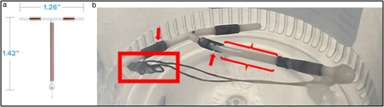
Actinomyces spp. has been shown to form biofilms when exposed to copper, possibly enhancing its degradation. Fragmentation and migration of the copper coil on an intrauterine device (IUD) is rare, but the concomitant presence of Actinomyces spp. may increase its incidence. We present the first case of a fragmented copper IUD within its lifespan of 10 years, with documented Actinomyces genitourinary tract colonization in a premenopausal woman.
Keywords: Actinomyces; intrauterine device.
Plain language summary
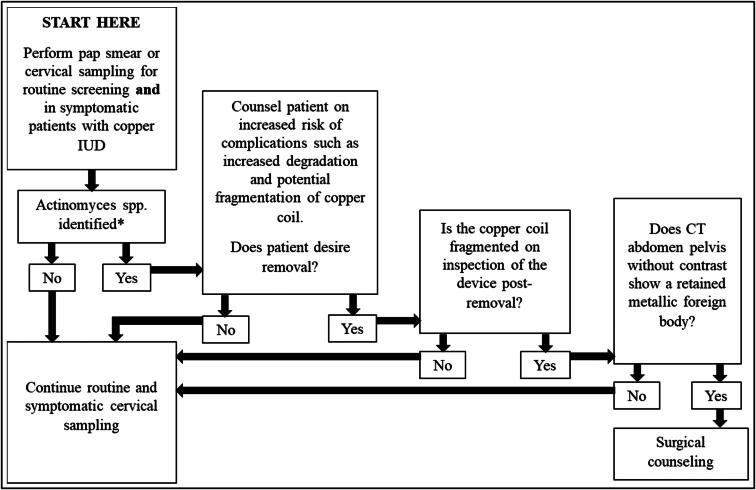
Actinomyces spp. are more prevalent in the urogenital tract of women with IUDs due to the local tissue injury from the device inside the uterus, which creates an anaerobic environment that allows this organism to flourish.Actinomyces spp. can form biofilms when exposed to copper, potentially enhancing its degradation, leading to fragmentation.Patients with a copper IUD and Actinomyces identified on routine or symptomatic cervical sampling should be counseled regarding potential enhanced copper degradation, fragmentation, and migration of the copper coil.
Copyright © 2024 Baylor University Medical Center.
Conflict of interest statement
The planners and faculty for this activity have no relevant financial relationships or funding to disclose. The patient consented to publication of this case report.
Figures
Similar articles
-
The effect of a copper intra-uterine contraceptive device on the microbial ecology of the female genital tract.J Med Microbiol. 1988 Apr;25(4):245-51. doi: 10.1099/00222615-25-4-245. J Med Microbiol. 1988. PMID: 3357191
-
Pelvic actinomycosis and usage of intrauterine contraceptive devices.Yale J Biol Med. 1982 Sep-Dec;55(5-6):453-61. Yale J Biol Med. 1982. PMID: 7183022 Free PMC article.
-
Incidence of actinomycosis associated with intrauterine devices.J Reprod Med. 1994 Aug;39(8):585-7. J Reprod Med. 1994. PMID: 7996521
-
The pathology of intra-uterine contraceptive devices.Curr Top Pathol. 1994;86:307-30. doi: 10.1007/978-3-642-76846-0_8. Curr Top Pathol. 1994. PMID: 8162713 Review.
-
Clinicopathological problems of the local tissue effect of the copper-releasing intrauterine contraceptive device (IUD). I. General characteristics of the copper-containing IUD (clinical study).Acta Chir Hung. 1989;30(2):129-32. Acta Chir Hung. 1989. PMID: 2669431 Review.
Cited by
-
Fragmentation of an In-Date Copper Intrauterine Device: An Unusual Complication.Cureus. 2024 Nov 9;16(11):e73344. doi: 10.7759/cureus.73344. eCollection 2024 Nov. Cureus. 2024. PMID: 39659323 Free PMC article.
References
-
- Patai K, Balogh I, Szarvas Z.. Clinicopathological problems of the local tissue effect of the copper-releasing intrauterine contraceptive device (IUD). I. General characteristics of the copper-containing IUD (clinical study). Acta Chir Hung. 1989;30(2):129–132. - PubMed
-
- Berthou J, Chrétien FC, Driguez PA.. Dégradation in utero des DIU au cuivre en fonction du temps. Le phénomène de corrosion métallique. Etude au microscope électronique à balayage [Degradation of copper IUDs in utero. The process of metallic corrosion. A scanning electron microscope study]. Gynecol Obstet Fertil. 2003;31(1):29–42. doi:10.1016/s1297-9589(02)00013-9. - DOI - PubMed
Publication types
LinkOut - more resources
Full Text Sources
Miscellaneous