. 2024 Dec;44(12):1422-1426.
doi: 10.1002/cac2.12623. Epub 2024 Oct 23.

IL2-mediated modulation of small extracellular vesicles secretion and PD-L1 expression: a novel perspective for neutralizing immune suppression within cancer cells

Affiliations

IL2-mediated modulation of small extracellular vesicles secretion and PD-L1 expression: a novel perspective for neutralizing immune suppression within cancer cells

Soojeong Noh et al. Cancer Commun (Lond). 2024 Dec.
No abstract available


Conflict of interest statement

The authors declare no competing interests.

Figures
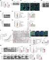
FIGURE 1
FIGURE 1
IL2 potently reduced sEV secretion and expression of both cPD‐L1 and ePD‐L1 in melanoma cells. (A‐B) B16F1 and B16F10 cells were incubated with 10 IU/mL IL2 for 48 h. (A) The number of sEVs secreted by B16F1 and B16F10 cells was measured by NTA (n = 3). (B) Protein expression of Rab27a in B16F1 and B16F10 cells was measured by western blotting. (C) Representative immunofluorescence images of cPD‐L1 (green) in mouse melanoma cells. Scale bars: 50 µm. DAPI (blue) indicates cell nuclei. Cells treated with PBS were used as a control (Con). (D) Expression of ePD‐L1 from B16F1 and B16F10 cells after 48 h of treatment with PBS or 10 IU/mL IL2. Protein expression of ePD‐L1 and sEV markers (ALIX, CD63, and TSG101) in cancer cell‐derived sEVs was analyzed by western blotting. (E‐F) B16F10 cells were treated with 10 IU/mL IL2 for 48 h, after IL2RB or IL2RG was blocked by probing with neutralizing antibodies, e.g. αIL2RB and αIL2RG for 1 h. (E) Protein expression of Rab27a and cPD‐L1 in cells was detected by western blotting. (F) Representative flow cytometric plots (left) and quantification (right) of ePD‐L1 expression (n = 4). (G‐H) B16F10 cells were co‐treated with 10 IU/mL IL2 and signaling inhibitors (PD98059, MEK inhibitor; LY294002, PI3K inhibitor; PIMOZIDE, STAT5 inhibitor) for 48 h. Protein expression of Rab27a and cPD‐L1 was determined by western blotting (G). Representative flow cytometric plots (left) and quantification (right) of ePD‐L1 expression (n = 4) (H). (I) B16F10‐luc‐g5 cells (WT), IL2‐T, or Thrombopoietin(TPO)‐T were transplanted subcutaneously into C57BL/6 mice. TPO‐T was used as a control. When the tumor reached a size of 30 mm3, the WT‐transplanted mice were injected intratumorally with PBS or IL2 (50,000 IU/mice) seven times every 2 days. Tumor volume of allografts in C57BL/6 mice (n = 11/group). (J) Representative IHC images of Rab27a (upper panel) and cPD‐L1 (lower panel) expression in tumor tissue. Scale bars: 100 µm (zoomed‐out) and 25 µm (zoomed‐in). (K) Representative immunocytochemistry (ICC) images (green: Rab27a, red: cPD‐L1, blue: DAPI). Scale bars: 100 µm. DAPI indicates cell nuclei. (L) Level of ePD‐L1 isolated from mouse plasma was verified by flow cytometry. Representative flow cytometric plots (left) and quantification (right) for ePD‐L1 expression (n = 3−5). (M) SK‐MEL‐28 xenograft models were established in CAnN.Cg‐Foxn1nu/Crl mice. When tumor size reached 30 mm3, PBS or IL2 (50,000 IU/mice) was injected intratumorally daily. Left: Representative western blot bands indicating protein expression of Rab27a and cPD‐L1 in mouse tumor tissues. Middle: Quantification of western blot bands to determine the ratio of Rab27a to β‐actin using Image J (n = 5). Right: Quantification of the western blot bands to determine the ratio of cPD‐L1 to β‐actin using Image J (n = 5). (N) Level of ePD‐L1 isolated from mouse plasma was measured by flow cytometry. Representative flow cytometric plots (left) and quantification (right) for ePD‐L1 expression (n = 11). (O) Protein expression of Rab27a and cPD‐L1 in lung cancer cells from seven lung tumor patients after 48 h of treatment with PBS or 10 IU/mL IL2 was detected by western blotting. Left: Representative western blot bands indicating protein expression of Rab27a and cPD‐L1. Middle: Quantification of the western blot bands to determine the ratio of Rab27a to β‐actin. Right: Quantification of the western blot bands to determine the ratio of cPD‐L1 to β‐actin using Image J. The p values of unpaired two‐tailed Student's t test in (A, M, N, O), one‐way ANOVA with Holm–Sidak's multiple comparisons test in (F, H, L) and two‐way ANOVA with Sidak's multiple comparisons test in (I): *P < 0.05; **P < 0.01; ***P < 0.001; ****P < 0.0001; ns, not significant. Representative data from triplicate experiments. Data are shown as mean ± SEM. Abbreviations: cPD‐L1, cellular programmed death‐ligand 1; ePD‐L1, exosomal programmed death‐ligand 1; ICC, immunocytochemistry; IHC, immunohistochemistry; IL2, interleukin‐2; sEV, small extracellular vesicle; WT, wide type.

References

    1. Becker A, Thakur BK, Weiss JM, Kim HS, Peinado H, Lyden D. Extracellular Vesicles in Cancer: Cell‐to‐Cell Mediators of Metastasis. Cancer Cell. 2016;30:836‐48. - PMC - PubMed
    1. Gong L, Yan Q, Zhang Y, Fang X, Liu B, Guan X. Cancer cell reprogramming: a promising therapy converting malignancy to benignity. Cancer Commun. 2019;39:48. - PMC - PubMed
    1. Blank C, Gajewski TF, Mackensen A. Interaction of PD‐L1 on tumor cells with PD‐1 on tumor‐specific T cells as a mechanism of immune evasion: implications for tumor immunotherapy. Cancer Immunol Immunother. 2005;54:307‐14. - PMC - PubMed
    1. Chen G, Huang AC, Zhang W, Zhang G, Wu M, Xu W, et al. Exosomal PD‐L1 contributes to immunosuppression and is associated with anti‐PD‐1 response. Nature. 2018;560:382‐6. - PMC - PubMed
    1. Pardoll DM. The blockade of immune checkpoints in cancer immunotherapy. Nat Rev Cancer. 2012;12:252‐64. - PMC - PubMed