. 2025 Feb;67(2):321-329.
doi: 10.1007/s00234-024-03486-w. Epub 2024 Oct 23.

Visualization of cortical neoangiogenesis after combined revascularization surgery in moyamoya disease using silent MRA

Affiliations

Visualization of cortical neoangiogenesis after combined revascularization surgery in moyamoya disease using silent MRA

Tomoaki Suzuki et al. Neuroradiology. 2025 Feb.

Abstract

Purpose: To investigate postsurgical indirect cortical neoangiogenesis in patients with moyamoya disease (MMD) using silent magnetic resonance angiography (MRA).

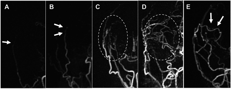
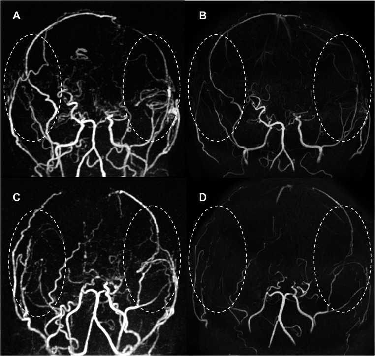
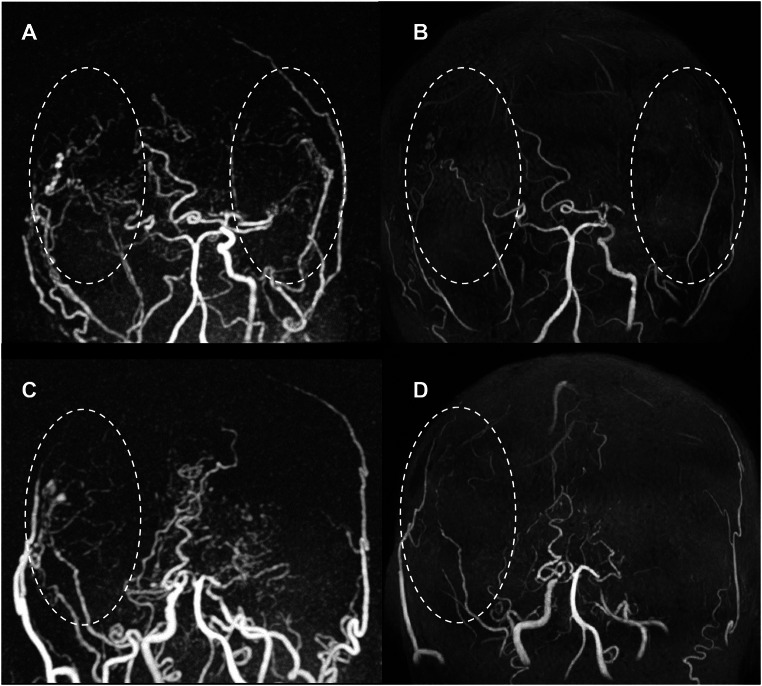
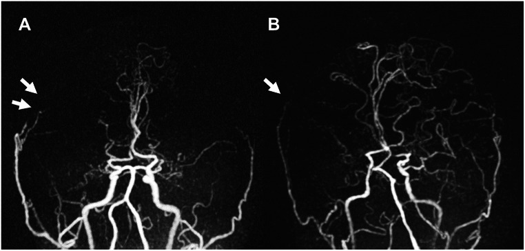
Methods: We studied 44 patients with MMD (63 hemispheres) who were previously revascularized with combined bypass surgery (23 and 40 hemispheres in pediatric and adult patients, respectively). They underwent follow-up for postoperative bypass patency using time-of-flight (TOF)-MRA and silent MRA between January 2022 and December 2023. The mean duration from surgery to MRA was 8.5 years (range, 1.2-22.3 years). Two observers independently rated the revascularization as follows: 0 (near-complete signal loss or no signal); 1, poor (slightly visible donor arteries); 2, good (acceptable revascularization around the brain surface); and 3, excellent (good quality of revascularization with perfusion from the cortical surface into the middle cerebral artery).

Results: Silent MRA visualized indirect bypass significantly better than TOF-MRA (2.6 ± 0.7 and 1.4 ± 0.8) (P < 0.01). In silent MRA, the mean score of indirect bypass was significantly higher than that of direct bypass (2.6 ± 0.7 and 1.7 ± 1.0; P < 0.01) and indicated good indirect bypass development in both children and adults (91.3% and 85.0%; score ≥ 2). Children exhibited a higher rate of excellent indirect bypass patency than adults (73.9% and 55.0%; score 3). Poor bypass development in indirect bypass (8 hemispheres, mean age: 35.5 ± 17.5 years, mean follow-up period: 11.3 years) was significantly observed in male patients (P < 0.01).

Conclusion: Silent MRA enables better precision in postsurgical visualization of indirect cortical neoangiogenesis during long-term follow-up and reveals indirect bypass development even in adult patients.

Keywords: Cortical neoangiogenesis; Indirect bypass; Moyamoya disease; Silent MRA; UTE-MRA.


Conflict of interest statement

Declarations. Ethics approval: This study was approved by the Ethics Committee of Niigata University Medical and Dental Hospital (approval number: 2019 − 0187). Informed consent: Patient consent was waived because of the retrospective nature of the study and the analysis used anonymous clinical data. Disclosures: None.

Figures

Fig. 1
Fig. 1
Grading score of postsurgical bypass patency on a four-point scale. (A) Grade 0, not visible (near-complete signal loss or no signal of donor arteries; white arrow); (B) grade 1, poor (slightly visible donor arteries; white arrows); (C) grade 2, good (acceptable revascularization around the brain surface; white dotted circle); (D) grade 3 in indirect bypass, excellent (good quality of revascularization with perfusion from the cortical surface into the middle cerebral artery; white dotted circle); and (E) grade 3 in direct bypass (white arrows)
Fig. 2
Fig. 2
Representative MRA images of pediatric patients in long-term follow-up. A 14-year-old girl, who had previously suffered from transient ischemic attack, was treated with combined direct and indirect bypass (superficial temporal artery [STA]–middle cerebral artery [MCA] single anastomosis with encephalomyosynangiosis [EMS] and encephalodurosynangiosis [EDS]) for both hemispheres. Fourteen years after bypass surgery, follow-up (A) silent MRA and (B) TOF-MRA were performed. (A) Based on silent MRA, the scores of the two observers were 2.5 for direct bypass and 3 for indirect bypass on the right side and 1 for direct bypass and 3 for indirect bypass on the left side (white dotted circles). (B) Based on TOF-MRA, the scores of the two observers were 1 for direct bypass and 1.5 for indirect bypass on the right side and 2 for direct bypass and 2 for indirect bypass on the left side (white dotted circles). An 8-year-old girl, who had previously suffered from epilepsy, was treated with combined direct and indirect bypass (STA-MCA single anastomosis with EMS and EDS) for both hemispheres. Twenty-two years after bypass surgery, follow-up (C) silent MRA and (D) TOF-MRA were performed. (C) Based on silent MRA, the scores of the two observers were 2.5 for direct bypass and 3 for indirect bypass on the right side and 1 for direct bypass and 3 for indirect bypass on the left side (white dotted circles). (D) Based on TOF-MRA, the scores of the two observers were 1.5 for direct bypass and 0.5 for indirect bypass on the right side and 1.5 for direct bypass and 0 for indirect bypass on the left side (white dotted circles)
Fig. 3
Fig. 3
Representative MRA images of adult patients in long-term follow-up. A 51-year-old woman, who had previously suffered from cerebral infarction, was treated with combined direct and indirect bypass (superficial temporal artery [STA]–middle cerebral artery [MCA] single anastomosis with encephalomyosynangiosis [EMS] and encephalodurosynangiosis [EDS]) for both hemispheres. Fourteen years after bypass surgery, follow-up (A) silent MRA and (B) TOF-MRA were performed. (A) Based on silent MRA, the scores of the two observers were 1 for direct bypass and 3 for indirect bypass on the right side and 1 for direct bypass and 3 for indirect bypass on the left side (white dotted circles). (B) Based on TOF-MRA, the scores of the two observers were 0.5 for direct bypass and 2.5 for indirect bypass on the right side and 1 for direct bypass and 0 for indirect bypass on the left side (white dotted circles). A 41-year-old woman, who had previously suffered from cerebral infarction, was treated with combined direct and indirect bypass (right STA-MCA single anastomosis with EMS and EDS). Twenty-two years after bypass surgery, follow-up (C) silent MRA and (D) TOF-MRA were performed. (C) Based on silent MRA, the mean scores of the two observers were 2.5 for direct bypass and 3 for indirect bypass (white dotted circle). (D) Based on TOF-MRA, the scores of the two observers were 1.5 for direct bypass and 1 for indirect bypass (white dotted circle)
Fig. 4
Fig. 4
Representative MRA images of patients with poor indirect bypass development. (A) A 49-year-old man, who had previously suffered from cerebral infarction, was treated with combined direct and indirect bypass (right superficial temporal artery [STA]–middle cerebral artery [MCA] single anastomosis with encephalomyosynangiosis [EMS] and encephalodurosynangiosis [EDS]). Two years after bypass surgery, follow-up silent magnetic resonance angiography (MRA) was performed. The mean scores of the two observers were 1.5 for direct bypass and 0 for indirect bypass (white arrows). (B) A 39-year-old man, who had previously suffered from cerebral infarction, was treated with combined direct and indirect bypass (STA-MCA single anastomosis with EMS and EDS). Seventeen years after bypass surgery, follow-up silent MRA was performed. The scores of the two observers were 0 for direct bypass and 0 for indirect bypass on the right side (white arrow) and 2.5 for direct bypass and 1 for indirect bypass on the left side

Similar articles

Cited by

References

    1. Suzuki J, Takaku A (1969) Cerebrovascular moyamoya disease. Disease showing abnormal net-like vessels in base of brain. Arch Neurol 20:288–299. 10.1001/archneur.1969.00480090076012 - PubMed
    1. Kuroda S, Houkin K (2008) Moyamoya disease: current concepts and future perspectives. Lancet Neurol 7:1056–1066. 10.1016/S1474-4422(08)70240-0 - PubMed
    1. Macyszyn L, Attiah M, Ma TS, Ali Z, Faught R, Hossain A, Man K, Patel H, Sobota R, Zager EL, Stein SC (2017) Direct versus indirect revascularization procedures for moyamoya disease: a comparative effectiveness study. J Neurosurg 126:1523–1529. 10.3171/2015.8.JNS15504 - PubMed
    1. Park S-E, Kim J-S, Park EK, Shim K-W, Kim D-S (2018) Direct versus indirect revascularization in the treatment of moyamoya disease. J Neurosurg 129:480–489. 10.3171/2017.5.JNS17353 - PubMed
    1. Ge P, Zhang Q, Ye X, Liu X, Deng X, Wang J, Wang R, Zhang Y, Zhang D, Zhao J (2020) Postoperative collateral formation after indirect bypass for hemorrhagic moyamoya disease. BMC Neurol 20:28. 10.1186/s12883-020-1612-z - PMC - PubMed

LinkOut - more resources